medical and biological sciences - Collegium Medicum - Uniwersytet ...
medical and biological sciences - Collegium Medicum - Uniwersytet ...
medical and biological sciences - Collegium Medicum - Uniwersytet ...

You also want an ePaper? Increase the reach of your titles
YUMPU automatically turns print PDFs into web optimized ePapers that Google loves.
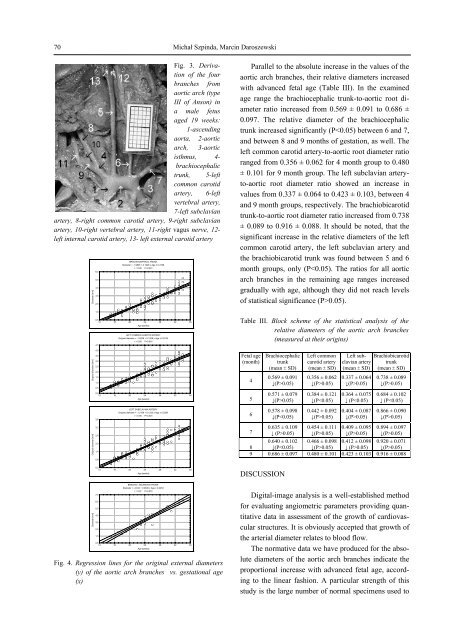
70Michał Szpinda, Marcin DaroszewskiFig. 3. Derivationof the fourbranches fromaortic arch (typeIII of Anson) ina male fetusaged 19 weeks:1-ascendingaorta, 2-aorticarch, 3-aorticisthmus, 4-brachiocephalictrunk, 5-leftcommon carotidartery, 6-leftvertebral artery,7-left subclavianartery, 8-right common carotid artery, 9-right subclavianartery, 10-right vertebral artery, 11-right vagus nerve, 12-left internal carotid artery, 13- left external carotid arteryDiameter [mm]Original diameter [mm]Original diameter [mm]Diameter [mm]6,55,54,53,52,51,5BRACHIOCEPHALIC TRUNKDiameter = -1.9835 + 0.1948 x Age ± 0.3728r = 0.95 P0.05)0.364 ± 0.075↓ (P0.05)0.409 ± 0.095↓(P>0.05)Brachiobicarotidtrunk(mean ± SD)0.738 ± 0.089↓(P>0.05)0.684 ± 0.102↓ (P0.05)0.894 ± 0.097↓(P>0.05)70.640 ± 0.102 0.466 ± 0.098 0.412 ± 0.098 0.920 ± 0.0718 ↓(P0.05) ↓ (P>0.05) ↓(P>0.05)9 0.686 ± 0.097 0.480 ± 0.101 0.423 ± 0.103 0.916 ± 0.088DISCUSSIONDigital-image analysis is a well-established methodfor evaluating angiometric parameters providing quantitativedata in assessment of the growth of cardiovascularstructures. It is obviously accepted that growth ofthe arterial diameter relates to blood flow.The normative data we have produced for the absolutediameters of the aortic arch branches indicate theproportional increase with advanced fetal age, accordingto the linear fashion. A particular strength of thisstudy is the large number of normal specimens used to