medical and biological sciences - Collegium Medicum - Uniwersytet ...
medical and biological sciences - Collegium Medicum - Uniwersytet ...
medical and biological sciences - Collegium Medicum - Uniwersytet ...
You also want an ePaper? Increase the reach of your titles
YUMPU automatically turns print PDFs into web optimized ePapers that Google loves.
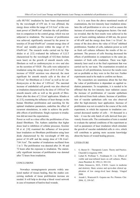
Effect of Low Level Laser Therapy <strong>and</strong> High Intensity Laser Therapy on endothelial cell proliferation in vitro... 83cells HUVEC irradiation by laser beam characterizedby the wavelength of 670 nm. It was affirmed, theenergy doses within the range of 2-8 J/cm 2. at the constantpower of 20mW/cm 2 stimulate the cell proliferationin comparison to the control group, which was notsubjected to irradiation. The increase of proliferationwas achieved, significantly stressed by the power intensitiesof 20 <strong>and</strong> 65mW/cm 2. constant energy dose of8J/cm 2 <strong>and</strong> variable power within the range of 10-65mW/cm 2 . The research works carried out by Kipshidzeet al. [12] evaluated the influence of LLLTcharacterized by the wavelength of 632.8nm (heliumneon laser) on the growth of smooth muscle cells,fibrolasts as well as cardiomyocytes in vitro <strong>and</strong> alsoon the secretion of VEGF. The cells were subjected toirradiation using the energy doses of 01-6.3J/cm 2 . Theincrease of VEGF secretion was observed; the mostsignificant for smooth muscle cells at the dose of0.5J/cm 2. for fibroblasts at 2.1J/cm 2 as well as for cardiomyocytesat the dose of 1.05J/cm 2 . The maximalincrease of endothelial cell growth was also observedat irradiation characterized by the dose of 1.05J/cm 2 forsmooth muscle cells as well as the growth of fibroblastsafter the dose of 2.1J/cm 2 application. Khadra etal. [13], examining the influence of laser therapy on thehuman fibroblast proliferation <strong>and</strong> searching for theoptimal irradiation parameters, underline the effect ofrecurrent stimulations, in order to achieve the profitableeffects of proliferation. Single exposure to irradiationdid not meet the expectations.Power as well as a dose affect the proliferation of irradiatedfibroblasts. The Authors underline that higherdoses lead to inhibition of cellular processes. KreislerM et al. [18] examined the influence of low-powerlaser irradiation on fibroblasts proliferation using laserdiode characterized by the wavelength of 808 nm,power of 10 mW <strong>and</strong> the energy dose of 1.96 J/cm 2 -7.84 J/cm 2 . The number of irradiations oscillated from1 to 3. The proliferation was denoted after 24. 48 <strong>and</strong>72 hours after the exposure to irradiation. The statisticallysignificant increase of proliferation was denotedafter 72 hours from irradiation.CONCLUSIONSNowadays neoangiogenesis presents widely analyzedmarker of tissues healing, thus the studies concerningmethods of tissue proliferation increase areneeded. It will help to develop a blood vessel networkin case of treatment of hard-to-heal wounds.As it is seen from the above mentioned results ofexaminations, the low-intensity laser irradiation stimulatesthe cell proliferation in vitro as well as causes theincrease of secretion of angiogenous factors. Our studiesrevealed, that the best results were achieved in thecase of lasers emitting radiation of 660 nm, the powerof 40 mW <strong>and</strong> the energy dose from 1 to 5J/cm 2 . Increaseof the dose above the value of 5J/cm 2 inhibitedproliferation. Number of cells, radiation power as wellas fresh cell cultures influence the results of the experiment.The process of cells freezing decreased theeffects of proliferation in comparison to the same parametersof fresh cells irradiation. There was highintensitylaser used in at the final experiment that wascarried out by our team. The examination revealed anincrease of cell proliferation, however the results werenot so profitable as they were in the first test. Furtherexperiments need to be made to confirm our theses.The researchers are unanimous about the fact thatthe value of proliferation is dependant on applied radiationparameters. To recapitulate, it is needed to beaffirmed that the low-intensity laser radiation causesthe increase of proliferation of vascular epitheliumcells derived from fresh cultures. Increase of proliferationof vascular epithelium cells was also observedafter the high-intensity laser application. Increase ofproliferation was not revealed in the course of the sixthexperiment, in which the exposure to irradiation concerneddecreased number of cells – 10 thous<strong>and</strong> in ahole – it was the only batch of cells derived from previouslyfrozen cells. The continuation of tests is neededto evaluate the optimal conditions of the experiment aswell as parameters of laser irradiation which stimulatethe growth of vascular endothelial cells in vitro, whichwill contribute to gaining more accurate knowledgeabout the basics of wound healing process .LITERATURE1. G. Baxter D. : Therapeutic Lasers. Theory <strong>and</strong> Practice.Churchill Livingstone 1994.2. Lubart R, Wollman Y., Freidmann H. i in.: Effects ofvisible <strong>and</strong> near-infrared lasers on cell cultures. PhotochemPhotobiol. B. 1992,12, 305-310.3. Zlatko Simunovic, M.D., F.M.H.: Lasers in medicine<strong>and</strong> dentistry: Basic science <strong>and</strong> up-to-date clinical applicationof low energy-level laser therapy. Vitagraf2000.4. Arendt J., Waniczek D.: Gojenie ran. Prz. Piśmien. Chir.2004, 12, 359-367.


