atw International Journal for Nuclear Power | 04.2020
Title atw - International Journal for Nuclear Power | 04.2020 Description Ever since its first issue in 1956, the atw – International Journal for Nuclear Power has been a publisher of specialist articles, background reports, interviews and news about developments and trends from all important sectors of nuclear energy, nuclear technology and the energy industry. Internationally current and competent, the professional journal atw is a valuable source of information. www.nucmag.com
Title
atw - International Journal for Nuclear Power | 04.2020
Description
Ever since its first issue in 1956, the atw – International Journal for Nuclear Power has been a publisher of specialist articles, background reports, interviews and news about developments and trends from all important sectors of nuclear energy, nuclear technology and the energy industry. Internationally current and competent, the professional journal atw is a valuable source of information.
www.nucmag.com

Create successful ePaper yourself
Turn your PDF publications into a flip-book with our unique Google optimized e-Paper software.
<strong>atw</strong> Vol. 65 (2020) | Issue 4 ı April<br />
RESEARCH AND INNOVATION 228<br />
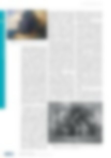
| Fig. 5.<br />
A landscape painting with a large oak tree, assigned to a 19 th century artist.<br />
available with additional options like<br />
automated scanning of the surface of<br />
the painting and automated recording<br />
of the data from the investigated<br />
object. Further optimizations are<br />
precise, very small irradiation spots in<br />
so- called Micro-XRF instruments<br />
using collimating X-ray lenses. The<br />
advantage is at hand: The area under<br />
investigation is much better characterised<br />
and the imaging resolution is<br />
higher, while the scanning over the<br />
whole surface reveals a complete<br />
image of the elemental distribution.<br />
These X-ray images seem much better<br />
achievable than to expend high ef<strong>for</strong>ts<br />
<strong>for</strong> neutron activation radiographies,<br />
although it should be reminded that<br />
low-Z elements are difficult or not at<br />
all detectable in XRF because of the<br />
attenuation of their characteristic<br />
X-rays in the paint layers. Modern<br />
scanning Micro-XRF instruments are<br />
operated in investigations of important<br />
art objects like in the actual<br />
analysis prior to the restoration of<br />
Rembrandt´s painting “Night Watch”<br />
of the Rijksmuseum in Amsterdam<br />
[12].<br />
In the following example measured<br />
at TÜV NORD – the case of a landscape<br />
painting from the 19 th Century<br />
– the power of X-ray radiography<br />
combined with XRF analysis is<br />
demonstrated (Figures 5 and 6). The<br />
painting, showing several traces of old<br />
damages, had been restored earlier<br />
and by this occasion had been relined<br />
(this means, a second canvas was<br />
glued on the backside). The XRF<br />
results from the elements in the paint<br />
layers were consistent <strong>for</strong> the pigments<br />
expected like iron (pigment<br />
Siena or Umber) and manganese<br />
( pigment Umber). The detection of<br />
chromium (pigment Chrome Green<br />
from the beginning of the 19 th century,<br />
or the improved version of<br />
hydrated chromium oxide, which<br />
became known as Viridian Green<br />
during the 1860’s) allows to date the<br />
painting into the 19 th century. Be<strong>for</strong>e<br />
the introduction of these chromium<br />
pigments, green colors in paintings<br />
were mostly based on copper pigments<br />
like Azurite, Malachite or<br />
Verdigris, however, the element<br />
copper was not detected in the XRF<br />
spectra of this painting. It is worth<br />
noting, that similar XRF pigment<br />
analysis is well suited to detect fake<br />
paintings if the <strong>for</strong>ger used modern<br />
pigments like Titanium White (available<br />
since ca. 1920) or various<br />
Cadmium pigments (not available<br />
be<strong>for</strong>e the very late 19 th Century), as it<br />
was discovered in a number of cases of<br />
art <strong>for</strong>gery. The most prominent case<br />
might be the <strong>for</strong>ger Wolfgang Beltracci<br />
who specialized on fakes of Expressionism<br />
painters from around 1900<br />
and probably sold hundreds of fakes<br />
into the art market. In one of his<br />
<strong>for</strong>ged paintings of the famous painter<br />
Campendonk, the pigment Titanium<br />
White was measured in a technical<br />
investigation with XRF, which proved<br />
that the painting was a fake and<br />
thereby uncovered one of the largest<br />
cases of art <strong>for</strong>gery.<br />
In the landscape painting of<br />
Figure 5, surprisingly a high amount<br />
of mercury (an element, which is<br />
umambiguously assigned to the red<br />
pigment Cinnabar) was detected. The<br />
idea, that Cinnabar could have been<br />
used in one of the paint layers below<br />
the visible surface was at hand. By<br />
means of the digital X-ray radiography<br />
of the painting, it was discovered that<br />
the artist had used an older canvas<br />
with a mythological or religious<br />
figural scenery <strong>for</strong> his work (see<br />
Figure 6) and that the overpainted<br />
and hidden composition is well<br />
consistent with the use of Cinnabar<br />
pigment, which often occurs in (red)<br />
clothes of the figures.<br />
As it was stated above, X-ray<br />
radiography is not limited to paintings<br />
and this is shown in the following case<br />
[13]. Near the village of Schortens<br />
in Friesland, a part of North-West-<br />
Germany, a large cemetery field from<br />
the early middle-ages had been<br />
discovered in the 1970’s. The archaeological<br />
findings, attributed to a period<br />
from the 5 th to the 12 th Century,<br />
consisted of cinerary urns including a<br />
variety of corroded metallic artefacts<br />
and are conserved in museums today<br />
(Landesmuseum Natur und Mensch<br />
Oldenburg and Landesmuseum<br />
Hannover). However, most of the<br />
metallic artefacts were never investigated,<br />
although the presumed keys,<br />
buckles, knives or even swords given<br />
to the graves of the dead would reveal<br />
additional knowledge about the life in<br />
these early populations of North-<br />
West- Germany, be<strong>for</strong>e and during the<br />
Christianisation phase. The ef<strong>for</strong>t to<br />
restore such objects is very high and<br />
restoration is under the risk of<br />
damaging the objects during the<br />
delicate procedures involved. Hence,<br />
a non-destructive imaging by X-rays<br />
was chosen in order to reveal first<br />
insight into the corroded pieces and<br />
prepare a decision basis <strong>for</strong> later<br />
restoring.<br />
As an example <strong>for</strong> the results,<br />
Figure 7 shows the amazing pictures<br />
of a so-called needle tubule, which<br />
was an important part of women´s<br />
equipment in the early middle ages,<br />
| Fig. 6.<br />
X-ray radiography (30 kV, 4 mAs, © TÜV NORD EnSys) of the painting from Fig.5, showing the older and<br />
overpainted composition as well as structural in<strong>for</strong>mation.<br />
Research and Innovation<br />
Radiation in Art and Cultural Heritage ı Frank Meissner and Andrea Denker