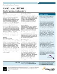
Medical Applications User Guide (pdf) - Freescale Semiconductor
Medical Applications User Guide (pdf) - Freescale Semiconductor
Medical Applications User Guide (pdf) - Freescale Semiconductor

Create successful ePaper yourself
Turn your PDF publications into a flip-book with our unique Google optimized e-Paper software.
18.2<br />
Ultrasound<br />
Ultrasound is a non-invasive medical imaging<br />
technique used to visualize muscles, tendons,<br />
pathological lesions and many internal organs<br />
and other structures. It plays an important role<br />
during prenatal care and is commonly used as<br />
a diagnostic tool.<br />
One of the most common uses of ultrasound<br />
is for fetal monitoring. Ultrasound uses sound<br />
waves to create images of a fetus inside<br />
a uterus. Because it uses sound waves<br />
instead of radiation, ultrasound is safer than<br />
X-rays. Gradually, ultrasound has become an<br />
increasingly important part of prenatal care,<br />
providing information that can help the doctor<br />
to plan the monitoring of a pregnant woman,<br />
thus improving the chances of successful<br />
pregnancy.<br />
18.3<br />
How Ultrasound Works<br />
Ultrasound is based on bouncing sound<br />
waves into the body of the developing fetus.<br />
The echoes produced by these waves are<br />
converted into a picture called a sonogram,<br />
which appears on a monitor. This technique is<br />
also often referred to as sonography or sonar.<br />
Propagation and reflection rules that govern<br />
electric signals are also applied to ultrasound.<br />
A transmission line must be terminated in its<br />
characteristic impedance to avoid reflections.<br />
In the equation below, acoustic impedance<br />
Z is a fundamental property of matter and<br />
is related to the density ρ and the velocity<br />
of sound v : Z = ρv. The fraction of energy<br />
R refracted at the normal interface of two<br />
different tissue types is:<br />
R = (Z 2 -Z 1 )<br />
(Z 2 +Z 1 )<br />
2<br />
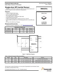
Figure 18-1: Ultrasound General Block Diagram<br />
Ultrasound<br />
<strong>Freescale</strong> Technology Optional<br />
18.4<br />
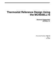
Transducer<br />
The transducer is the element that converts<br />
electrical signals into ultrasound waves. It<br />
consists of a set of transmitter and receiver<br />
transducers arranged in a linear array. A<br />
unique transducer is explained in Chapter<br />
16.6, Fetal Heart Rate Monitor. Pulse trains<br />
are sent by transmitter transducers, and<br />
receiver transducers receive bounced waves.<br />
The operating frequency for this kind of device<br />
is from 5 MHz to 8 MHz.<br />
<strong>Medical</strong> Imaging<br />
HV Pulse<br />
Transducer DAC<br />
TX Beamformer<br />
Generator<br />
Tx/Rx<br />
Switches<br />
LNA<br />
Signal Conditioning<br />
The blocks needed for signal conditioning/<br />
pulse generator blocks are shown in<br />
Figure 18-3.<br />
18.5<br />
Multiplexer for Tx/Rx<br />
Transducers<br />
This block may be implemented using analog<br />
gates controlled by the MCU/MPU. This<br />
allows the use of transducers as transmitters,<br />
and later the ability to switch the multiplexer<br />
to use as receivers. Multiplexing reduces the<br />
freescale .com/medical 95<br />
VGA<br />
CW (Analog)<br />
Beamformer<br />
AAF<br />
Power<br />
Management<br />
Keypad<br />
DAC<br />
ADC<br />
ADC<br />
<strong>User</strong> Interface<br />
Spectral<br />
Doppler<br />
Processing<br />
(D Mode)<br />
Display Memory Audio<br />
Output<br />
Figure 17-2: 18-2: Ultrasound Transducer Diagram Diagram<br />
TX<br />
RX<br />
TX<br />
RX<br />
TX<br />
RX<br />
TX<br />
RX<br />
RX Beamformer<br />
RF<br />
Demodulation<br />
B-Mode<br />
Processing<br />
Scan<br />
Conversion<br />
USB<br />
Patient<br />
Beamforming<br />
Control<br />
DSP/DSC<br />
Color<br />
Doppler<br />
(PW)<br />
Processing<br />
(F Mode)<br />
Wireless<br />
Comm