Medical Applications User Guide (pdf) - Freescale Semiconductor
Medical Applications User Guide (pdf) - Freescale Semiconductor
Medical Applications User Guide (pdf) - Freescale Semiconductor

Create successful ePaper yourself
Turn your PDF publications into a flip-book with our unique Google optimized e-Paper software.
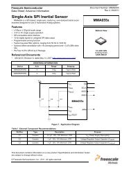
<strong>Medical</strong> Imaging<br />
number of connections needed, because the<br />
transducers array can range from eight to<br />
more than 256.<br />
18.6<br />
Instrumentation Amplifier<br />
and Variable Gain<br />
Amplifier<br />
Ultrasonic wave energy sent though a<br />
patient’s body is very attenuated by multiple<br />
factors (absorbing, attenuation due to the<br />
medium, inverse square law, etc.). Before<br />
processing information, the instrumentation<br />
amplifier conditions the signal to adequate<br />
levels and eliminates common-mode noise.<br />
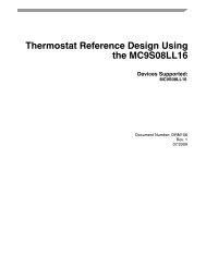
A variable gain amplifier is used due to<br />
exponential attenuation of the bounced<br />
waves. Applying an exponential gain reduces<br />
the effect of the attenuation. Figure 18-4<br />
shows the behavior of this element.<br />
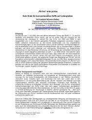
Figure 18-5 shows a simple analog<br />
implementation of the circuit (left side). At the<br />
right side, a block diagram of a control system<br />
is shown. This can be implemented by an<br />
MPU using software.<br />
18.7<br />
Beamformer<br />
A beamformer is a device that directs waves<br />
in a specific direction by means of algorithms<br />
that control the transducer array to form<br />
a wave front that generates constructive<br />
interference. This is used to generate the<br />
sweep required to build the image to be<br />
shown. Figure 18-7 is a diagram of the<br />
direction of propagation of waves controlled<br />
by a beamformer.<br />
18.8<br />
Ultrasound Software<br />
Library<br />
The ultrasound software library produces an<br />
ultrasound image from a beamforming signal.<br />
The beam is stored in the memory and passes<br />
through the ultrasound library algorithms to<br />
generate an output image with the specified<br />
height and width.<br />
Figure Figure 17-3: 18-3: Ultrasound Ultrasound Probe Probe Block Block Diagram Diagram<br />
Transducer Array<br />
Figure 18-4: Variable Gain Amplifier Function<br />
Figure 17-4: Variable Gain Amplifier Function<br />
Amplitude<br />
Multiplexer<br />
for TX/RX<br />
Transducers<br />
Instrumentation<br />
Amplifier<br />
High-Voltage<br />
TX Amplifier<br />
Figure 18-5: Analog Implementation of Variable Gain Amplifier<br />
96 <strong>Medical</strong> <strong>Applications</strong> <strong>User</strong> <strong>Guide</strong><br />
Gain<br />
Fixed<br />
Gain<br />
Variable Gain<br />
Amplifier<br />
High-Speed<br />
DAC<br />
Time<br />
High-Speed<br />
High-Resolution<br />
ADC<br />
TX<br />
Beamformer<br />
Amplitude<br />
RX<br />
Beamformer<br />
Beamformer<br />
Control<br />
System<br />
Time<br />
To DSP Blocks