HYPERTONIA ÉS NEPHROLOGIA - eLitMed.hu
HYPERTONIA ÉS NEPHROLOGIA - eLitMed.hu
HYPERTONIA ÉS NEPHROLOGIA - eLitMed.hu

Create successful ePaper yourself
Turn your PDF publications into a flip-book with our unique Google optimized e-Paper software.
16 MARKUS KETTELER <strong>HYPERTONIA</strong> <strong>ÉS</strong> <strong>NEPHROLOGIA</strong><br />
implicated that elevated phosphate<br />
concentrations may additionally induce<br />
a phenotypic switch of vascular<br />
smooth muscle cells into osteoblastlike<br />
cells. Jono et al. demonstrated in<br />
cultured vascular smooth muscle cells<br />
(VSCM) that an elevation of the phosphate<br />
concentration in the culture medium<br />
from 1.4 to 2.0 mmol/l indeed<br />
caused hydroxyapatite formation, but<br />
that this effect was clearly dependent<br />
on a rise in the intracellular phosphate<br />
concentration (27). Moreover, this<br />
group observed the formation of matrix<br />
vesicles, an upregulation of the<br />
osteoblast transcription factor cbfa-1<br />
and de novo expression of bonerelated<br />
proteins including alkaline<br />
phosphatase, osteopontin and osteocalcin<br />
by VSCM under high phosphate<br />
culture conditions, indicating<br />
an osteogenic dedifferentiation of this<br />
cell type (27). These data, together<br />
with the immunohistochemical detection<br />
of cbfa-1, alkaline phosphatase<br />
and osteopontin in calcified epigastric<br />
arteries by Sharon Moe’s group (28,<br />
29), suggest that hyperphosphatemiadependent<br />
vascular calcification is an<br />
active cellular process in uremic patients.<br />
In view of these observations,<br />
any therapeutic means to decrease<br />
phosphate load and to prevent<br />
hyperphosphatemia in order to suppress<br />
extraosseous calcifications are<br />
certainly of major importance.<br />
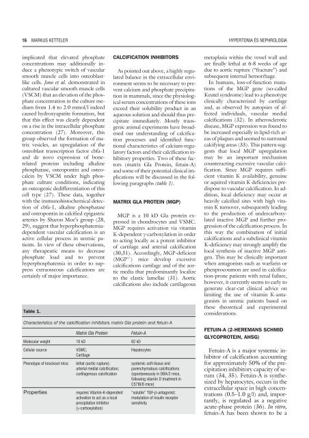
Table 1.<br />
CALCIFICATION INHIBITORS<br />
As pointed out above, a highly regulated<br />
balance in the extracellular environment<br />
seems to be necessary to prevent<br />
calcium and phosphate precipitation<br />
in mammals, since the physiological<br />
serum concentrations of these ions<br />
exceed their solubility product in an<br />
aqueous solution and should t<strong>hu</strong>s precipitate<br />
immediately. Mostly transgenic<br />
animal experiments have broadened<br />
our understanding of calcification<br />
processes and identified functional<br />
characteristics of calcium-regulatory<br />
factors and their calcification inhibitory<br />
properties. Two of these factors<br />
(matrix Gla Protein, fetuin-A)<br />
and some of their potential clinical implications<br />
will be discussed in the following<br />
paragraphs (table 1).<br />
MATRIX GLA PROTEIN (MGP)<br />
MGP is a 10 kD Gla protein expressed<br />
in chondrocytes and VSMC.<br />
MGP requires activation via vitamin<br />
K-dependent -carboxylation in order<br />
to acting locally as a potent inhibitor<br />
of cartilage and arterial calcification<br />
(30,31). Accordingly, MGP-deficient<br />
(MGP -/- ) mice develop excessive<br />
calcifications cartilage and of the aortic<br />
media that predominantly localize<br />
to the elastic lamellae (31). Aortic<br />
calcifications also include cartilageous<br />
Characteristics of the calcification inhibitors matrix Gla protein and fetuin-A<br />
Matrix Gla Protein<br />
Fetuin-A<br />
Molecular weight 10 kD 62 kD<br />
Cellular source<br />
Phenotype of knockout mice<br />
Properties<br />
VSMC;<br />
Cartilage<br />
lethal (aortic rupture);<br />
arterial medial calcification;<br />
cartilagenous calcification<br />
requires Vitamin-K-dependent<br />
activation to act as a local<br />
precipitation inhibitor<br />
(-carboxylation)<br />
Hepatocytes<br />
systemic soft-tissue and<br />
parenchymatous calcifications<br />
(spontaneously in DBA/2 mice,<br />
following vitamin D treatment in<br />
C57Bl/6 mice)<br />
“soluble” TGF--antagonist;<br />
modulation of insulin receptor<br />
sensitivity<br />
metaplasia within the vessel wall and<br />
are finally lethal at 6-8 weeks of age<br />
due to aortic rupture (“fracture”) and<br />
subsequent internal hemorrhage.<br />
In <strong>hu</strong>mans, loss-of-function mutations<br />
of the MGP gene (so-called<br />
Keutel syndrome) lead to a phenotype<br />
clinically characterized by cartilage<br />
and, as observed by autopsies of affected<br />
individuals, vascular medial<br />
calcifications (32). In atherosclerotic<br />
disease, MGP expression was found to<br />
be increased especially in lipid-rich areas<br />
of plaques and seemed to surround<br />
calcifying areas (33). This pattern suggests<br />
that local MGP upregulation<br />
may be an important mechanism<br />
counteracting excessive vascular calcification.<br />
Since MGP requires sufficient<br />
vitamin K availability, genuine<br />
or aquired vitamin K deficiencies predispose<br />
to vascular calcification. In addition,<br />
local deficiency may occur at<br />
heavily calcified sites with high vitamin<br />
K turnover, subsequently leading<br />
to the production of undercarboxylated<br />
inactive MGP and further progression<br />
of the calcification process. In<br />
this way the combination of initial<br />
calcifications and a subclinical vitamin<br />
K-deficiency may strongly amplify the<br />
local synthesis of inactive MGP antigen.<br />
This may be clinically important<br />
when antagonists such as warfarin or<br />
phenprocoumon are used in calcification-prone<br />
patients with renal failure,<br />
however, it currently seems to early to<br />
generate clear-cut clinical advice on<br />
limiting the use of vitamin K-antagonists<br />
in uremic patients based on<br />
these theoretical and experimental<br />
considerations.<br />
FETUIN-A (2-HEREMANS SCHMID<br />
GLYCOPROTEIN, AHSG)<br />
Fetuin-A is a major systemic inhibitor<br />
of calcification accounting<br />
for approximately 50% of the precipitation<br />
inhibitory capacity of serum<br />
(34, 35). Fetuin-A is synthesized<br />
by hepatocytes, occurs in the<br />
extracellular space in high concentrations<br />
(0.5–1.0 g/l) and, importantly,<br />
is regulated as a negative<br />
acute-phase protein (36). In vitro,<br />
fetuin-A has been shown to be a