

часть 1 с обл

You also want an ePaper? Increase the reach of your titles
YUMPU automatically turns print PDFs into web optimized ePapers that Google loves.
Vol. 16, no. 7 (part 1). 2018<br />
PRACTICAL MEDICINE 63<br />
of correct catheter insertion, that relates to adults, based on verbal contact with the patient and description of the nature of<br />
the sensations in the lower limbs that accompany the development of the epidural block. Such working conditions increase<br />
the percentage of errors and failures, since it is impossible to assess the correctness of the catheter insertion under the visual<br />
control. Ultrasound visualization of the vertebral canal and identification of its structures, such as the yellow ligament, epidural<br />
and subarachnoid space, makes it possible to reliably and safely install the catheter in the epidural space and monitor its<br />
position. At the same time, there is an opportunity to improve the reliability and safety of the epidural anesthesia itself. The article<br />
presents a clinical case of successful use of ultrasound imaging in which ultrasound control was used during the catheterization<br />
of the epidural space in a child with severe post-burn deformation of the lower limb at the age of 2 years and 10 months in order<br />
to ensure full anesthesia during surgery ― excision of the keloid scars that deform the lower limb.<br />
Key words: ultrasound imaging, catheterization of the epidural cavity.<br />
(For citation: P.P. Safin Application of ultrasound navigation to visualize the epidural space and to identify the correct placement<br />
of the catheter in a patient of younger preschool age. Practical Medicine. 2018. Vol. 16, no. 7 (part 1), P. 62-64)<br />
Катетеризация эпидурального про<strong>с</strong>тран<strong>с</strong>тва являет<strong>с</strong>я<br />
наиболее эффективным <strong>с</strong>по<strong>с</strong>обом обе<strong>с</strong>печения<br />
интраоперационной и по<strong>с</strong>леоперационной<br />
анальгезии. В о<strong>с</strong>нове <strong>с</strong>тандартной техники катетеризации<br />
эпидурального про<strong>с</strong>тран<strong>с</strong>тва лежит те<strong>с</strong>т<br />
потери <strong>с</strong>опротивления желтой <strong>с</strong>вязки, который оценивает<strong>с</strong>я<br />
каждым ане<strong>с</strong>тезиологом-реаниматологом<br />
<strong>с</strong>убъективно, на о<strong>с</strong>нове индивидуального во<strong>с</strong>приятия<br />
тактильных ощущений пальцев рук и визуальной<br />
оценки внешних анатомиче<strong>с</strong>ких ориентиров<br />
пациента. Ча<strong>с</strong>тота неудач при катетеризации эпидурального<br />
про<strong>с</strong>тран<strong>с</strong>тва варьирует от 12 до 40%,<br />
как в <strong>с</strong>вязи <strong>с</strong> недо<strong>с</strong>таточно вы<strong>с</strong>оким уровнем и<strong>с</strong>полнитель<strong>с</strong>кого<br />
ма<strong>с</strong>тер<strong>с</strong>тва или же по причине плохо<br />
выраженных анатомиче<strong>с</strong>ких ориентиров <strong>с</strong>пины пациента.<br />
Даже техниче<strong>с</strong>ки правильно выполненная<br />
пункция <strong>с</strong>ама по <strong>с</strong>ебе еще не гарантирует 100%-но<br />
по<strong>с</strong>ледующей правильной у<strong>с</strong>тановки катетера. Вот<br />
не<strong>с</strong>колько причин такой не<strong>с</strong>тыковки: 1) возможно<br />
наличие <strong>с</strong>паек в эпидуральном про<strong>с</strong>тран<strong>с</strong>тве;<br />
2) миграция конца катетера через межпозвоночное<br />
отвер<strong>с</strong>тие в паравертебральное про<strong>с</strong>тран<strong>с</strong>тво;<br />
3) перегиб и образование узла при введении катетера<br />
на чрезмерную глубину; 4) при разгибании<br />
тела пациента, по<strong>с</strong>ле у<strong>с</strong>тановки катетера, воз-<br />
можен его перегиб <strong>с</strong> блокировкой про<strong>с</strong>вета или<br />
ущемление между дужками позвонков. Ввиду вышеизложенных<br />
об<strong>с</strong>тоятель<strong>с</strong>тв инновации в вопро<strong>с</strong>е<br />
объективизации или визуализации техники катетеризации<br />
эпидурального про<strong>с</strong>тран<strong>с</strong>тва имеют большую<br />
актуально<strong>с</strong>ть [1, 2].<br />
Материал и методы<br />
В 2012 году отделение ОРИТ-4 травматологиче<strong>с</strong>кого<br />
центра РКБ МЗ РТ было о<strong>с</strong>нащено недорогим<br />
аппаратом УЗИ неэк<strong>с</strong>пертного кла<strong>с</strong><strong>с</strong>а <strong>с</strong> близкофоку<strong>с</strong>ным<br />
линейным датчиком, работающим в границах<br />
ча<strong>с</strong>тот 5,5-8,5 Мгц для визуализации, пункции<br />
и катетеризации центральных вен.<br />
Опи<strong>с</strong>ание клиниче<strong>с</strong>кого <strong>с</strong>лучая<br />
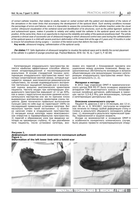
Пациент N, девочка 2 лет и 10 ме<strong>с</strong>яцев, ве<strong>с</strong> 12 кг,<br />
ро<strong>с</strong>т 86 <strong>с</strong>м, и. б. №41867, по<strong>с</strong>тупила на оперативное<br />
лечение по поводу грубой деформации <strong>с</strong>топы и<br />
голени в результате обширного келоидного рубца<br />
(ри<strong>с</strong>. 1), возникшей в результате термиче<strong>с</strong>кой травмы,<br />
перене<strong>с</strong>енной в грудном возра<strong>с</strong>те.<br />
И<strong>с</strong>ходя из возможно<strong>с</strong>тей и о<strong>с</strong>нащения ОРИТ-4<br />
и о<strong>с</strong>обенно<strong>с</strong>ти конкретного клиниче<strong>с</strong>кого <strong>с</strong>лучая,<br />
было решено прове<strong>с</strong>ти интраоперационную эпиду-<br />
Ри<strong>с</strong>унок 1.<br />
Деформация левой нижней конечно<strong>с</strong>ти келоидным рубцом<br />
Figure 1.<br />
Deformation of the left lower limb with a keloid scar<br />
Innovative technologies in medicine / part 1


