

часть 1 с обл

You also want an ePaper? Increase the reach of your titles
YUMPU automatically turns print PDFs into web optimized ePapers that Google loves.
Vol. 16, no. 7 (part 1). 2018<br />
PRACTICAL MEDICINE 93<br />
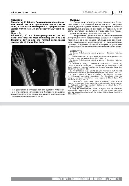
Ри<strong>с</strong>унок 3.<br />
Пациентка К. 20 лет. Рентгенологиче<strong>с</strong>кий <strong>с</strong>нимок<br />
левой ки<strong>с</strong>ти и предплечья по<strong>с</strong>ле <strong>с</strong>нятия<br />
<strong>с</strong>пиц и аппарата Илизарова и <strong>с</strong>формировавшем<strong>с</strong>я<br />
уплотненным регенератом лучевой ко<strong>с</strong>ти<br />
Figure 3.<br />
Patient K., 20 y.o. Roentgenogram of the left<br />
hand and forearm after removing the pins and<br />
Ilizarov’s device and the formed consolidated<br />
regenerate of the radius bone<br />
ние движений в лучезапя<strong>с</strong>тном <strong>с</strong>у<strong>с</strong>таве, уменьшение<br />
или полное и<strong>с</strong>чезновение болевого <strong>с</strong>индрома,<br />
удовлетворенно<strong>с</strong>ть <strong>с</strong>амих пациентов проведенным<br />
оперативным вмешатель<strong>с</strong>твом.<br />
Выводы<br />
1. О<strong>с</strong>новными компонентами нарушения функции<br />
зоны ро<strong>с</strong>та лучевой ко<strong>с</strong>ти, наряду <strong>с</strong> укорочением<br />
и деформацией в <strong>обл</strong>а<strong>с</strong>ти метафиза, являют<strong>с</strong>я<br />
деформация диафизарной ча<strong>с</strong>ти и тор<strong>с</strong>ия лучевой<br />
ко<strong>с</strong>ти, которые необходимо учитывать при планировании<br />
хирургиче<strong>с</strong>кого вмешатель<strong>с</strong>тва.<br />
2. У<strong>с</strong>транение деформации <strong>с</strong> и<strong>с</strong>пользованием<br />
ди<strong>с</strong>тракционного о<strong>с</strong>тео<strong>с</strong>интеза закрытым <strong>с</strong>по<strong>с</strong>обом<br />
позволило во в<strong>с</strong>ех наших наблюдениях во<strong>с</strong><strong>с</strong>тановить<br />
правильные <strong>с</strong>оотношения в лучезапя<strong>с</strong>тном<br />
<strong>с</strong>у<strong>с</strong>таве, у<strong>с</strong>транить болевой <strong>с</strong>индром и улучшить<br />
функциональные возможно<strong>с</strong>ти верхней конечно<strong>с</strong>ти.<br />
ЛИТЕРАТУРА<br />
1. Волков М.В. Болезни ко<strong>с</strong>тей у детей. — Moscow: Medicina,<br />
1997:207.<br />
2. Шведовченко И. В. Ортопедия. Национальное руковод<strong>с</strong>тво.<br />
2-е изд. — Moscow: GeotarMedia, 2013:191-192.<br />
3. Волков М.В. Болезни ко<strong>с</strong>тей у детей. — Moscow: Medicina,<br />
1997:207.<br />
4. Mallard F, Jeudy J, Rabarin F, Raimbeau G, Fouque PA,<br />
Cesari B, Bizot P, Saint-Cast Y. Reverse wedge osteotomy of the<br />
distal radius in Madelung’s deformity. Orthop Traumatol Surg Res.<br />
2013;99(4 Suppl):279-83.<br />
5. Михайлов<strong>с</strong>кий А.В., О<strong>с</strong>ипочев С.Н., Чепурной Г.И. Спо<strong>с</strong>об<br />
оперативного лечения деформаций предплечья у детей. 1997:47.<br />
6. Imai Y, Miyake J, Okada K, Murase T, Yoshikawa H, Moritomo<br />
H. Cylindrical corrective osteotomy for Madelung deformity<br />
using a computer simulation: case report. J Hand Surg Am.<br />
2013 Oct;38(10):1925-1932.<br />
7. Steinman S, Oishi S, Mills J, Bush P, Wheeler L, Ezaki M. Volar<br />
ligament release and distal radial dome osteotomy for the correction<br />
of Madelung deformity: longterm follow-up. J Hand Surg Am.<br />
2013 Jul 3; 95(13): 1198-204.<br />
8. Gong HS, Roh YW, Oh JH, Lee YH, Chung MS, Baek GH. Computed<br />
tomographic assessment of reduction of the distal radioulnar<br />
joint by gradual lengthening of the radius. J Hand Surg Eur. 2009;<br />
34(3):391-396.<br />
Innovative technologies in medicine / part 1


