

X-Ray Fluorescence Analytical Techniques - CNSTN : Centre ...
X-Ray Fluorescence Analytical Techniques - CNSTN : Centre ...
X-Ray Fluorescence Analytical Techniques - CNSTN : Centre ...

You also want an ePaper? Increase the reach of your titles
YUMPU automatically turns print PDFs into web optimized ePapers that Google loves.
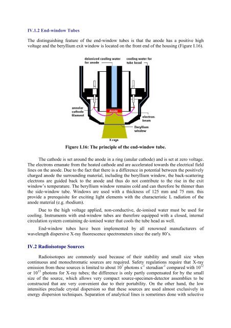
IV.1.2 End-window Tubes<br />
The distinguishing feature of the end-window tubes is that the anode has a positive high<br />
voltage and the beryllium exit window is located on the front end of the housing (Figure I.16).<br />
Figure I.16: The principle of the end-window tube.<br />
The cathode is set around the anode in a ring (anular cathode) and is set at zero voltage.<br />
The electrons emanate from the heated cathode and are accelerated towards the electrical field<br />
lines on the anode. Due to the fact that there is a difference in potential between the positively<br />
charged anode the surrounding material, including the beryllium window, the back-scattering<br />
electrons are guided back to the anode and thus do not contribute to the rise in the exit<br />
window’s temperature. The beryllium window remains cold and can therefore be thinner than<br />
the side-window tube. Windows are used with a thickness of 125 mm and 75 mm. this<br />
provide a prerequisite for exciting light elements with the characteristic L radiation of the<br />
anode material (e.g. rhodium).<br />
Due to the high voltage applied, non-conductive, de-ionised water must be used for<br />
cooling. Instruments with end-window tubes are therefore equipped with a closed, internal<br />
circulation system containing de-ionised water that cools the tube head as well.<br />
End-window tubes have been implemented by all renowned manufacturers of<br />
wavelength dispersive X-ray fluorescence spectrometers since the early 80’s.<br />
IV.2 Radioisotope Sources<br />
Radioisotopes are commonly used because of their stability and small size when<br />
continuous and monochromatic sources are required. Safety regulations require that X-ray<br />
emission from these sources is limited to about 10 7 photons s -1 steradian -1 compared with 10 12<br />
or 10 13 photons for X-ray tubes; the difference is only partly compensated for by the small<br />
size of the source, which allows very compact source-specimen-detector assemblies to be<br />
constructed that are very convenient due to their portability. On the other hand, the low<br />
intensities preclude crystal dispersion so that these sources are used almost exclusively in<br />
energy dispersion techniques. Separation of analytical lines is sometimes done with selective