

2012 Proceedings - International Tissue Elasticity Conference
2012 Proceedings - International Tissue Elasticity Conference
2012 Proceedings - International Tissue Elasticity Conference

Create successful ePaper yourself
Turn your PDF publications into a flip-book with our unique Google optimized e-Paper software.
037 ACQUISITON OF FREEHAND ELASTOGRAPHY VOLUMES IN BRAIN TUMOR SURGERY:<br />
PRELIMINARY RESULTS.<br />
Tormod Selbekk 1,2 , Frank Lindseth 1,2 , Geirmund Unsgård 2,3 .<br />
1 SINTEF, Trondheim, NORWAY; 2 The Norwegian University of Science and Technology,<br />
Trondheim, NORWAY; 3 St. Olav Hospital, Trondheim University Hospital, Trondheim, NORWAY.<br />
Background: Ultrasound imaging is an established method for intraoperative guidance and monitoring in<br />
surgery of brain tumors. Some commercial systems also offer the possibility of performing freehand<br />
acquisition of ultrasound image volumes. In such systems, an ultrasound scanner is integrated with a<br />
navigation system which enables spatial tracking of the ultrasound probe equipped with a position sensor<br />
device. The ultrasound volume is generated by tilting and translating the ultrasound probe. After acquisition<br />
the ultrasound image volume is stored and subsequently displayed on the navigation system. This enables<br />
navigation and simultaneous display of intraoperative ultrasound and preoperative images as, e.g., MRI.<br />
Several research groups have explored ultrasound elastography or strain imaging of brain tumors [1–3], and<br />
real time 2D ultrasound elastography images have also been compared with co–registered MR–images using a<br />
navigation system [4]. Elastography (strain magnitude) images of glial brain tumors have been reported to<br />
have higher image contrast than corresponding B–mode images and may, therefore, serve as a complement in<br />
distinguishing resectable cancerous tissue from normal brain tissue [5]. It can be speculated that navigation<br />
of surgical instruments based on ultrasound elastography image volumes could contribute to detecting and<br />
localizing residual tumor tissue during surgery and thereby increase the proportion of gross total resection.<br />
We present our initial experience with freehand acquisition of elastography image volumes in brain surgery<br />
and neuronavigation based on simultaneous display of intraoperative elastography, B–mode ultrasound and<br />
preoperative, registered Magnetic Resonance image volumes.<br />
Aims: The aim of the study was to investigate whether or not it is possible to acquire ultrasound<br />
elastography image volumes of brain tumors using a simple freehand technique.<br />
Methods: Data have been acquired using the Ultrasonix MDP scanner (Vancouver, BC, Canada) with a flat<br />
linear probe (L14–5/38) and the standard elastography module. The navigation system consisted of the<br />
in–house developed CustusX research navigation software and the Polaris tracking system (NDI, Ontario,<br />
Canada). So far, data have been acquired in two clinical cases, a glioblastoma and a meningioma.<br />
Results: It was possible to acquire ultrasound elastography volumes of the brain tumor in both cases.<br />
Various display techniques like volume rendering and image fusion of ultrasound elastography volumes,<br />
conventional ultrasound and preoperative MRI has been explored.<br />
Conclusions: The initial test shows that it is possible to acquire and use ultrasound elastography volumes<br />
for neuronavigation purposes. The method must be further developed and refined for further clinical use.<br />
Acknowledgements: This work was financed by the National Centre for Ultrasound and Image Guided Therapy at St.<br />
Olav’s Hospital, Trondheim, Norway. We acknowledge the work of the CustusX development team: Christian<br />
Askeland, Ole Vegard Solberg, Janne B. Bakeng, Lars E. Bø and Erlend Hofstad (all SINTEF).<br />
References:<br />
[1] Selbekk T, Bang J, Unsgaard G: Strain Processing of Intraoperative Ultrasound Images of Brain Tumours: Initial<br />
Results. Ultrasound in Medicine and Biology, 31 (1), pp. 45–51, 2005.<br />
[2] Scholz M, Lorenz A et al.: Current Status of intraoperative Real-Time Vibrography in Neurosurgery. Ultraschall<br />
Med, 28 (5), pp. 493–497, 2007.<br />
[3] Uff CE, Garcia L, et al.: Real Time Ultrasound Elastography in Neurosurgery. <strong>Proceedings</strong> of the IEEE<br />
<strong>International</strong> Ultrasonics Symposium, pp. 467–470, 2009.<br />
[4] Chakraborty A, Berry G et al.: Intra–Operative Ultrasound Elastography and Registered Magnetic Resonance<br />
Imaging of Brain Tumours: A Feasibility Study. Ultrasound, 14 (1), pp. 43–49, 2006.<br />
[5] Selbekk T, Brekken R, Indergaard M, Solheim O, Unsgård G: Comparison of Contrast in Brightness Mode and<br />
Strain Ultrasonography of Glial Brain Tumours. BMC Med Imaging, 12(11), <strong>2012</strong>.<br />
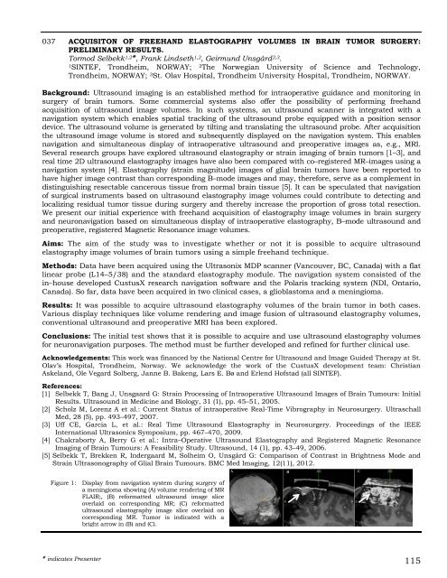
Figure 1: Display from navigation system during surgery of<br />
a meningioma showing (A) volume rendering of MR<br />
FLAIR;, (B) reformatted ultrasound image slice<br />
overlaid on corresponding MR; (C) reformatted<br />
ultrasound elastography image slice overlaid on<br />
corresponding MR. Tumor is indicated with a<br />
bright arrow in (B) and (C).<br />
indicates Presenter 115


