

2012 Proceedings - International Tissue Elasticity Conference
2012 Proceedings - International Tissue Elasticity Conference
2012 Proceedings - International Tissue Elasticity Conference

Create successful ePaper yourself
Turn your PDF publications into a flip-book with our unique Google optimized e-Paper software.
002 ANALYSIS OF THYROID NODULES VISCOELASTICITY WITH TIME STRAIN CURVES MAY<br />
SURPASS CLASSICAL QUALITATIVE AND SEMI–QUANTITATIVE EVALUATION.<br />
Rafal Z Slapa 1 , Bartosz Migda 1 , Wiesław S Jakubowski 1 , Jacek Bierca 2 ,<br />
Jadwiga Slowinska–Srzednicka 3 .<br />
1 Diagnostic Imaging Department, Medical University of Warsaw, Faculty of Medicine II with<br />
English and Physiotherapy Divisions, Warsaw, POLAND; 2 Surgery Department, Solec Hospital,<br />
Warsaw; POLAND; 3 Endocrinology Department, Center for Postgraduate Medical Education,<br />
Warsaw, POLAND.<br />
Background: It is assumed that elasticity is linear with deformation in the range of 1%. However,<br />
observation of time–strain curves of thyroid nodules subjected to free–hand strain elastography indicates<br />
both linear and non–linear portions of relaxation.<br />
Aims: The purpose of the study was to evaluate a new linear/non–linear approach for strain elastography<br />
of thyroid nodules based on the analysis of time strain curves and compare it with classical elasticity<br />
score and thyroid strain ratio methods.<br />
Methods: During 2009–2011, 67 patients scheduled for thyroidectomy (62 with multinodular goiter and<br />
5 with a single thyroid nodule) were evaluated with B–mode and power Doppler ultrasound of the whole<br />
thyroid and neck lymph nodes. During the ultrasound examination, 96 dominant nodules were examined<br />
with strain elastosonography with Aplio XG (Toshiba, Japan) with a linear 5–17MHz transducer. The<br />
stiffness of each thyroid nodule was evaluated on the pair of images with classical features of strain<br />
elastosonography qualitatively (with elasticity scores) and semi–quantitatively with thyroid tissue<br />
strain/nodule strain ratios with application of elasto Q (Toshiba, Japan). Moreover, a novel, original<br />
approach to elasticity data based on evaluation of time strain curves (elasto Q) was applied.<br />
Results: The incidence of thyroid cancer that was not known before our ultrasound examination<br />
(5 nodules) in our study shows that cancer is present in 9% of the 54 patients. Final diagnosis<br />
established 7 papillary carcinomas (6 in multinodular goiter), 4 follicular adenomas and 85 benign<br />
nodular goiter nodules. Classical elastographic analysis with elasticity score and elasticity ratio on<br />
statistical analysis did not show a significant difference between cancer and benign nodules<br />
(p–value=0,431 and 0,156, respectively). On linear/non–linear analysis of time strain curves, excellent<br />
differentiation (p=5.6x10⁻⁹) was possible with new parameter: the Relative Length of Non–Linear<br />
Relaxation (with threshold 0.5, sensitivity 100% and specificity 85.4%, area under ROC=0.975).<br />
Conclusions: (1) The incidence of thyroid cancer that was not known before our ultrasound examination<br />
in our study is comparable to the incidence of cancer in the general population of multinodular<br />
goiter patients even though the number of cancers in the study is small. (2) The analysis of linear<br />
and non–linear elastography data may greatly improve differential diagnosis of thyroid nodules. (3)<br />
Different timing for conversion from linear to non–linear relaxation between papillary thyroid cancer and<br />
benign thyroid nodules may indicate that viscosity may be the differential factor. (4) Further multicenter<br />
large scale studies evaluating the usefulness of linear/non–linear elastography phenomena in differential<br />
diagnostics of thyroid cancer are warranted. Studies involving evaluation of vioscoelasticity (e. g., shear<br />
wave spectroscopy) could be applied to investigate the model of thyroid nodules differentiation.<br />
Acknowledgements: I would like to thank Dr. Jeffrey Bamber and other lecturers at 10th ITEC 2011 in Arlington for<br />
inspiration. Supported by the Ministry of Science and Higher Education of Poland grant Nr N N402 476437, 2009–12.<br />
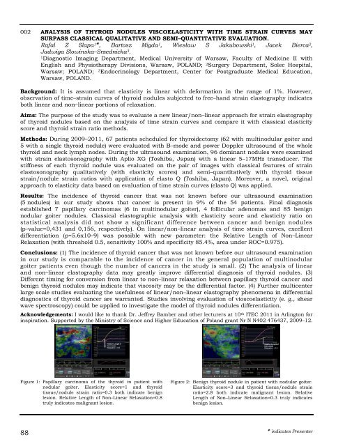
Figure 1: Papillary carcinoma of the thyroid in patient with<br />
nodular goiter. <strong>Elasticity</strong> score=1 and thyroid<br />
tissue/nodule strain ratio=0.3 both indicate benign<br />
lesion. Relative Length of Non–Linear Relaxation=0.8<br />
truly indicates malignant lesion.<br />
88<br />
Figure 2: Benign thyroid nodule in patient with nodular goiter.<br />
<strong>Elasticity</strong> score=3 and thyroid tissue/nodule strain<br />
ratio=2.8 both indicate malignant lesion. Relative<br />
Length of Non–Linear Relaxation=0.3 truly indicates<br />
benign lesion.<br />
indicates Presenter


