

2012 Proceedings - International Tissue Elasticity Conference
2012 Proceedings - International Tissue Elasticity Conference
2012 Proceedings - International Tissue Elasticity Conference

Create successful ePaper yourself
Turn your PDF publications into a flip-book with our unique Google optimized e-Paper software.
060 VISCOELASTICITY PARAMETERS OF HIFU ABLATED CANINE LIVER TISSUES EX VIVO.<br />
Danial Shahmirzadi 1 , Gary Y. Hou 1 and Elisa E. Konofagou 1 .<br />
1 Columbia University, VC 12–232, 622 West 168th Street, New York, NY, 10032, USA.<br />
Background: The feasibility of using elasticity imaging techniques for high–intensity–focused–ultrasound<br />
(HIFU) ablation monitoring and assessment has been previously demonstrated both in vitro and in vivo<br />
(e.g. [1,2]). Nonetheless, effective use of these techniques requires quantitative assessment of the changes<br />
in the mechanical properties of the ablated tissue during the treatment process. The present study<br />
focuses on the ablation effects on the liver tissue viscoelasticity ex vivo.<br />
Aims: To quantify the viscoelastic shear properties of thermally–ablated canine live tissue ex vivo.<br />
Methods: Canine liver specimens (n=6) were freshly excised immediately after sacrifice and kept<br />
immersed in de–ionized and degassed phosphate–buffered saline (PBS) at room temperature during the<br />
entire in vitro experimentation process. A therapeutic transducer (fcenter = 4.755MHz, fAM=25Hz,<br />
Riverside Research Inst, NY) was used to thermally ablate the liver test samples under 4 increasing<br />
HIFU energy levels of 36W×10s=360J (n=9; Group 1), 25W×30s=750J (n=15; Group 2),<br />
30W×30s=900J (n=18; Group 3), and 36W×30s=1080J (n=11; Group 4). After completion of the<br />
thermal ablation, the same test samples were tested regarding their viscoelastic parameters using a<br />
rheometer (TA Instrument, DE). The complex shear modulus (G*) and shear phase shift (δ) were<br />
obtained under a shear strain of ε=1% at a sweeping frequency range of f=0.1–10Hz. The<br />
measurements were also used to compute the tissue dynamic shear viscosity coefficient (η').<br />
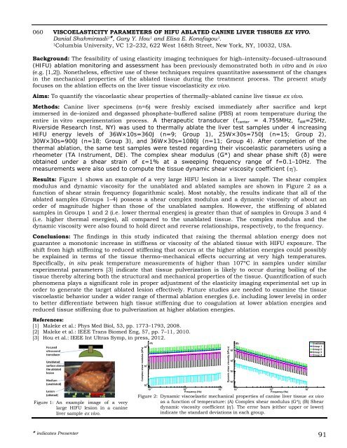
Results: Figure 1 shows an example of a very large HIFU lesion in a liver sample. The shear complex<br />
modulus and dynamic viscosity for the unablated and ablated samples are shown in Figure 2 as a<br />
function of shear strain frequency (logarithmic scale). Most notably, the results indicate that all of the<br />
ablated samples (Groups 1–4) possess a shear complex modulus and a dynamic viscosity of about an<br />
order of magnitude higher than those of the unablated samples. However, the stiffening of ablated<br />
samples in Groups 1 and 2 (i.e. lower thermal energies) is greater than that of samples in Groups 3 and 4<br />
(i.e. higher thermal energies), all compared to the unablated tissue. The complex modulus and the<br />
dynamic viscosity were also found to hold direct and reverse relationships, respectively, to the frequency.<br />
Conclusions: The findings in this study indicated that raising the thermal ablation energy does not<br />
guarantee a monotonic increase in stiffness or viscosity of the ablated tissue with HIFU exposure. The<br />
shift from high stiffening to reduced stiffening that occurs at the higher ablation energies could possibly<br />
be explained in terms of the tissue thermo–mechanical effects occurring at very high temperatures.<br />
Specifically, in situ peak temperature measurements of higher than 107ºC in samples under similar<br />
experimental parameters [3] indicate that tissue pulverization is likely to occur during boiling of the<br />
tissue thereby altering both the structural and mechanical properties of the tissue. Quantification of such<br />
phenomena plays a significant role in proper adjustment of the elasticity imaging experimental set up in<br />
order to generate the target ablated lesion effectively. Future studies are needed to examine the tissue<br />
viscoelastic behavior under a wider range of thermal ablation energies (i.e. including lower levels) in order<br />
to better differentiate between high tissue stiffening due to coagulation at lower ablation energies and<br />
reduced tissue stiffening due to pulverization at higher ablation energies.<br />
References:<br />
[1] Maleke et al.: Phys Med Biol, 53, pp. 1773–1793, 2008.<br />
[2] Maleke et al.: IEEE Trans Biomed Eng, 57, pp. 7–11, 2010.<br />
[3] Hou et al.: IEEE Int Ultras Symp, in press, <strong>2012</strong>.<br />
Figure 1: An example image of a very<br />
large HIFU lesion in a canine<br />
liver sample ex vivo.<br />
Complex shear modulus (kPa)<br />
10 2<br />
(A)<br />
10 1<br />
10 0<br />
10 -1<br />
10 0<br />
Frequency (Hz)<br />
indicates Presenter 91<br />
10 1<br />
Dynamic shear viscosity (kPa.s)<br />
10 1<br />
10 0<br />
10 -1<br />
10 -2<br />
(B)<br />
10 -1<br />
10 0<br />
Frequency (Hz)<br />
10 1<br />
Unablated<br />
Group 1<br />
Group 2<br />
Group 3<br />
Group 4<br />
Figure 2: Dynamic viscoelastic mechanical properties of canine liver tissue ex vivo<br />
as a function of temperature: (A) Complex shear modulus (G*); (B) Shear<br />
dynamic viscosity coefficient (η'). The error bars (either upper or lower)<br />
indicate the standard deviations in each group.


