

2012 Proceedings - International Tissue Elasticity Conference
2012 Proceedings - International Tissue Elasticity Conference
2012 Proceedings - International Tissue Elasticity Conference

You also want an ePaper? Increase the reach of your titles
YUMPU automatically turns print PDFs into web optimized ePapers that Google loves.
053 COMBINED ULTRASOUND AND BIOIMPEDANCE TO ASSESS TISSUE VISCO–ELASTICITY.<br />
Robert Dodde 1 , Grant Kruger 2 , Leo Koziol 1 , Jonathan Ophir 3 , William (Rick) F. Weitzel 1 .<br />
1 Internal Medicine, 2 Mechanical Engineering Departments, University of Michigan, Ann Arbor, MI<br />
48109, USA; 3 Diagnostic and Interventional Imaging Department, University of Texas Medical<br />
School, Houston, TX 77030, USA.<br />
Background: There is an enormous clinical need to manage disease states manifesting fluid overload by<br />
edema; these include chronic kidney disease (CKD), congestive heart failure (CHF), liver failure and<br />
lymphedema. Current practice entails serial physical examination and is crudely quantitative, rating<br />
edema on a scale of zero to 4+. In recent years, bioimpedance measurements have been shown to<br />
correlate with hydration status by tracking extracellular resistance over time. While helpful, confounding<br />
effects from cellular ionic content, particularly the sequestering of sodium throughout the body, hamper<br />
future evolution of this technique. Fortunately, complementary strategies based on ultrasound strain<br />
imaging can dynamically quantify and characterize highly non–linear and time dependent changes of a<br />
subsurface elastic medium under stress. Combining bio–impedance and ultrasound strain may offer a<br />
clinically useful and yet complete analysis of how edematous tissue responds to stimuli.<br />
Methods: Edematous tissue can be contrasted to normal tissue through their time–dependent response<br />
to a given stimulus. Physiologically, the removal of excess fluid from the tissue has a time–dependent<br />
component as does the reentry of fluid after stimuli are removed. In this manner, tissue can be viewed as<br />
a viscoelastic material exhibiting two unique phenomena: creep and relaxation. To test our hypothesis,<br />
both creep and relaxation tests were run on phantom materials. Creep was monitored using pulsed–wave<br />
ultrasound depth–dependent Doppler in a gelatin–based Metamucil phantom (to provide scattering) under<br />
50g, 100g and 200g constant loads. Relaxation was monitored using bioimpedance in cylindrical blocks<br />
of tofu (to mimic poroelastic fluid flow) under 10%, 20% and 30% constant strains. Data were fit to a<br />
standard linear solid (SLS) model for creep and relaxation.<br />
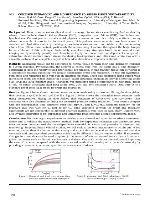
Results: Figure 1 below shows the creep measurements made using ultrasound. Fitting the data yielded<br />
time constants τε=12s±2s and τσ=113s±39s. Figure 2 below shows the relaxation measurements made<br />
using bioimpedance. Fitting the data yielded time constants of τεZ=2s±0.4s and τσZ=40s±8s. Time<br />
constants were also obtained by fitting the measured pressure during relaxation. These results compare<br />
with the bioimpedance time constants such that τεp=3τεp and τσp=0.75τσZ. Standard deviation for the<br />
pressure data was 0.7s for τεp and 3s for τσp. Time constants between the creep and relaxation<br />
experiments are not comparable as different phantom materials were used in each study. Current work<br />
also involves integration of bio–impedance and ultrasound phantoms into a single experimental model.<br />
Conclusions: We have begun experiments to develop a one dimensional quantitative edema assessment<br />
device and to validate the measurement method. Both bio–impedance relaxation and ultrasound creep<br />
measurements demonstrated the time–dependence essential for visco– and poro–elastic detection and<br />
monitoring. Targeting future clinical studies, we will seek to perform measurements within one to two<br />
minutes (rather than 8 minutes in this study) and expect this to depend on the force used and time<br />
constants and time dependent parameters which may be different in future human studies. If successful,<br />
changes in τ over time can be used to quantify the amount of edema removed from a tissue in order to<br />
guide treatments such as diuretic medications or dialysis. Our work continues directed toward improving<br />
the care of patients compared with the centuries old method of pressing on a patient’s extremity by<br />
providing a convenient, accurate, quantitative assessment of edema.<br />
120<br />
Figure 1: Measured and fitted creep strains. Gray<br />
areas are standard deviation limits (n=3).<br />
Figure 2: Measured and fitted admittance (1/|Z|)<br />
relaxation readings at varying strains.<br />
indicates Presenter


