

2012 Proceedings - International Tissue Elasticity Conference
2012 Proceedings - International Tissue Elasticity Conference
2012 Proceedings - International Tissue Elasticity Conference

Create successful ePaper yourself
Turn your PDF publications into a flip-book with our unique Google optimized e-Paper software.
016 HYPER–FREQUENCY VISCOELASTIC SPECTROSCOPY OF A VASCULAR–MIMICKING<br />
PHANTOM AND A PORCINE AORTA WITH THE RHEOSPECTRIS INSTRUMENT.<br />
Cédric Schmitt 1 , Anis Hadj Henni 1 , Simon LeFloc’h 2 , Jacques Ohayon 2 , Jonathan Vappou 3 , Guy Cloutier 4 .<br />
1 Rheolution Inc., Montréal, Québec, CANADA; 2 Laboratory TIMC–IMAG/DyCTiM, Université<br />
Joseph Fourier, Grenoble, FRANCE; 3 IMFS (FRE3240) / LSIIT (UMR 7005) CNRS – University of<br />
Strasbourg, Strasbourg, FRANCE; 4 Laboratory of Biorheology and Medical Ultrasonics, University<br />
of Montréal Hospital Research Center (CRCHUM), Montréal, Québec, CANADA.<br />
Background: Vascular elastography based on dynamic stress of arterial walls at frequencies typically up<br />
to 1500Hz is increasingly developed to monitor the mechanical properties of healthy and pathological<br />
vessels. However, available mechanical testing instruments can only measure viscoelastic properties up<br />
to 200Hz. Recently, a new instrument, RheoSpectrisTM (Rheolution Inc., Canada), has been introduced to<br />
perform viscoelastic spectroscopy of biomaterials between 10 and 1000Hz [1]. Thus, there is a great<br />
interest for using such an instrument to characterize vascular phantoms and tissues.<br />
Aims: The first objective was to measure the Young’s storage (E’) and loss (E’’) moduli of a vascular<br />
phantom material and a porcine aorta from 10 to 1000Hz with the hyper–frequency viscoelastic<br />
spectroscope RheoSpectrisTM C400 using a new beam geometry. The second objective was to compare, on<br />
phantom material, the viscoelasticity measurements with those obtained with DMA instruments.<br />
Methods: A standard silicone rubber (Platsil 71–10 silicone, Polytek Development Corp., USA) was first<br />
characterized using RheoSpectrisTM between 10 and 1000Hz with the bending modality (beam samples).<br />
For comparison, cylinders and plates made from the same material were tested using two DMA<br />
instruments: the Electroforce 3200 (Bose Corp, USA) using the compression modality and the Eplexor<br />
25N (GABO, Germany) using the compression and tensile modalities. Finally, beam–shaped samples<br />
(n=4) of a healthy porcine abdominal aorta were explanted and tested with RheoSpectrisTM .<br />
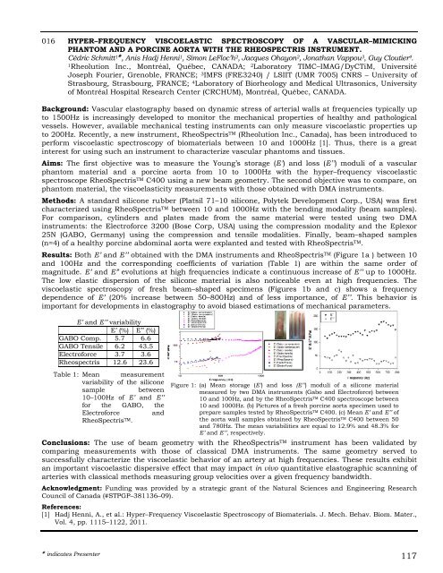
Results: Both E’ and E’’ obtained with the DMA instruments and RheoSpectrisTM (Figure 1a ) between 10<br />
and 100Hz and the corresponding coefficients of variation (Table 1) are within the same order of<br />
magnitude. E’ and E” evolutions at high frequencies indicate a continuous increase of E’’ up to 1000Hz.<br />
The low elastic dispersion of the silicone material is also noticeable even at high frequencies. The<br />
viscoelastic spectroscopy of fresh beam–shaped specimens (Figures 1b and c) shows a frequency<br />
dependence of E’ (20% increase between 50–800Hz) and of less importance, of E’’. This behavior is<br />
important for developments in elastography to avoid biased estimations of mechanical parameters.<br />
E’ and E’’ variability<br />
E’ (%) E’’ (%)<br />
GABO Comp. 5.7 6.6<br />
GABO Tensile 6.2 43.5<br />
Electroforce 3.7 3.6<br />
Rheospectris 12.6 23.6<br />
Table 1: Mean measurement<br />
variability of the silicone<br />
sample between<br />
10–100Hz of E’ and E’’<br />
for the GABO, the<br />
Electroforce and<br />
RheoSpectris TM.<br />
Figure 1: (a) Mean storage (E’) and loss (E’’) moduli of a silicone material<br />
measured by two DMA instruments (Gabo and Electroforce) between<br />
10 and 100Hz, and by the RheoSpectris TM C400 spectroscope between<br />
10 and 1000Hz. (b) Pictures of a fresh porcine aorta specimen used to<br />
prepare samples tested by RheoSpectris TM C400. (c) Mean E’ and E’’ of<br />
the aorta wall samples obtained by RheoSpectris TM C400 between 50<br />
and 780Hz. The mean variabilities are equal to 12.9% and 48.3% for<br />
E’ and E’’, respectively.<br />
Conclusions: The use of beam geometry with the RheoSpectrisTM instrument has been validated by<br />
comparing measurements with those of classical DMA instruments. The same geometry served to<br />
successfully characterize the viscoelastic behavior of an artery at high frequencies. These results exhibit<br />
an important viscoelastic dispersive effect that may impact in vivo quantitative elastographic scanning of<br />
arteries with classical methods measuring group velocities over a given frequency bandwidth.<br />
Acknowledgment: Funding was provided by a strategic grant of the Natural Sciences and Engineering Research<br />
Council of Canada (#STPGP–381136–09).<br />
References:<br />
[1] Hadj Henni, A., et al.: Hyper–Frequency Viscoelastic Spectroscopy of Biomaterials. J. Mech. Behav. Biom. Mater.,<br />
Vol. 4, pp. 1115–1122, 2011.<br />
indicates Presenter 117


