

2012 Proceedings - International Tissue Elasticity Conference
2012 Proceedings - International Tissue Elasticity Conference
2012 Proceedings - International Tissue Elasticity Conference

You also want an ePaper? Increase the reach of your titles
YUMPU automatically turns print PDFs into web optimized ePapers that Google loves.
032 ANALYSIS OF THE INFLUENCE OF INFLAMMATION, IRON AND WATER DIFFUSIVITY ON<br />
LIVER VISCOELASTIC PARAMETERS IN FIBROSIS.<br />
H Leitao 1,2,3 , S Doblas 1,2 , P Garteiser 1,2 , G D’Assignies 1,2 , F Mouri 1,2 , V Vilgrain 1,2 , R Sinkus 1,2 ,<br />
BE Van Beers 1,2 .<br />
1 INSERM, U773, F–75205, Paris, FRANCE; 2 University Paris Diderot, Sorbonne Paris Cité,<br />
UMRS773, F–75205, Paris, FRANCE; 3 University of Coimbra, Coimbra, PORTUGAL.<br />
Background: Increased liver steatosis, inflammation and iron are often observed in patients with liver<br />
fibrosis [1,2]. Moreover, the apparent diffusion coefficient (ADC) is associated with water content and the<br />
collagen matrix, and restricted diffusion is found with higher stages of fibrosis [3]. Yet, the influence of<br />
these factors on liver viscoelastic parameters determined by MR elastography is not fully understood.<br />
Aims: The purpose of our study was to assess the relationship between hepatic viscoelasticity and<br />
steatosis, inflammation, iron and ADC.<br />
Methods: All patient studies were performed under informed consent and within the agreement of local<br />
ethical guidelines. Thirty–seven patients with chronic viral hepatitis B (n=11) and C (n=26) underwent MR<br />
elastography (MRE), diffusion–weighted imaging (DWI) and multiecho imaging (ME) at 1.5T. Liver<br />
inflammation, fibrosis, inflammation and steatosis were assessed on percutaneous liver biopsies taken<br />
within a maximum delay of two months (mean 29 days). For MRE, we used a spin–echo EPI sequence<br />
(TR/TE=320ms/40ms, 4mm in–plane resolution, 3–directional encoding). Mechanical waves (50Hz) were<br />
coupled into the liver by an electromechanical actuator. DWI was performed with a spin echo EPI<br />
sequence (TR/TE=300/56, 4mm in–plane resolution, 11 b–values from 10 to 500s/mm 2 ). ME with an EPI<br />
readout (TR/TE=120/n×2.3ms; n=1;10) was used to quantify iron and fat. The following viscoelastic<br />
parameters were calculated by local inversion of the complex linear viscoelastic 3D wave equation:<br />
storage modulus, G’ (kPa), loss modulus G’’ (kPa), absolute value of the shear modulus, Gabs<br />
(Gabs 2 =G’ 2 +G’’ 2 ), propagation coefficient β (2π over the wavelength, mm -1 ) and attenuation, α (mm -1 ). The<br />
ADC was obtained by fitting a monoexponential function to the pixel intensities obtained at increasing<br />
diffusion sensitizing gradient strengths. Fat content was assessed by fitting separate monoexponential<br />
functions to the pixel intensities as a function of echo time on in–phase (water + fat) and out of phase<br />
(water – fat) echo images, subtracting the respective values predicted by the fit at zero echo time and<br />
dividing by two. The iron content was determined by mapping of the T2* MR relaxation coefficient<br />
according to the method exposed by Wood et al. [4]. Spearman rank correlation (r) and multiple<br />
regressions coefficients (RC) were calculated.<br />
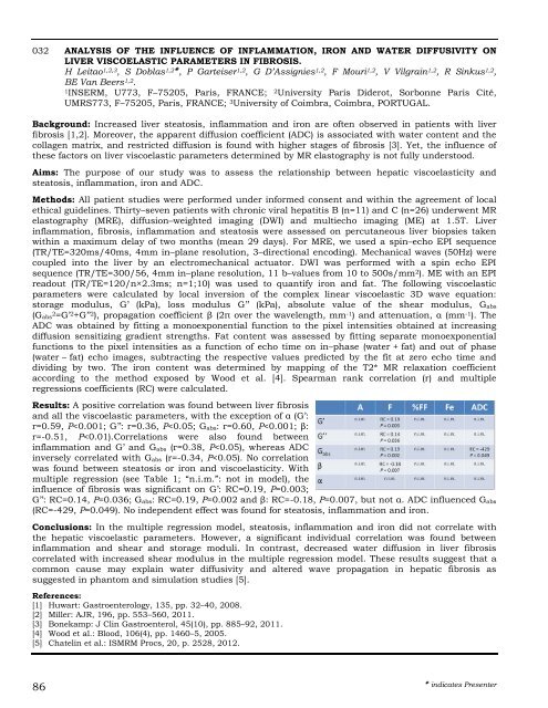
Results: A positive correlation was found between liver fibrosis<br />
and all the viscoelastic parameters, with the exception of α (G’:<br />
r=0.59, P


