Research Report 2010 2011 - Helmholtz-Zentrum für ...
Research Report 2010 2011 - Helmholtz-Zentrum für ...
Research Report 2010 2011 - Helmholtz-Zentrum für ...

You also want an ePaper? Increase the reach of your titles
YUMPU automatically turns print PDFs into web optimized ePapers that Google loves.
96 SCIENTIFIC REPORTS | Infection and Immunity | Infl ammation and Immunity<br />
03.5 Cell-death Mechanisms in Immunity<br />
PROJECT LEADER | Prof. Dr. Ingo Schmitz | <strong>Research</strong> Group Systems-oriented Immunology and Inflammation<br />
<strong>Research</strong> | isc09@helmholtz-hzi.de<br />
PROJECT MEMBERS | Michaela Annemann | Frida Ewald | Ralf Höcker | Dr. Yvonne Rauter | Marc Schuster | Tanja Telieps<br />
Types of cell death Multicellular organisms have evolved<br />
multiple cell-death mechanisms which are activated upon<br />
different stimuli and cell type-dependent to allow a finetuning<br />
of cellular responses to environmental conditions.<br />
Necrosis is characterized by swelling and rupture of the cells<br />
so that intracellular contents leak out of the cell and initiate<br />
an inflammatory immune response. During apoptosis, on<br />
the other hand, cells shrink and the integrity of the plasma<br />
membrane stays intact. Therefore, leakage of intracellular<br />
components does not take place and activation of the immune<br />
system is inhibited. Apoptosis plays an important role<br />
in the interplay between pathogens and the host cell. In<br />
this regard, immune cells may kill infected tissue cells by<br />
the induction of apoptosis. On the other hand, viruses and<br />
bacteria are able to inhibit host cell apoptosis to ensure their<br />
own survival and further spreading. Deregulation of apoptosis<br />
may lead to various diseases such as viral hepatitis (too<br />
much apoptosis) or cancer (too little apoptosis).<br />
Apoptotic signal transduction Two major apoptosis pathways<br />
exist referred to as the extrinsic and intrinsic pathway,<br />
respectively. The intrinsic pathway acts on the mitochondrium<br />
and is regulated by proteins of the Bcl-2 family. The<br />
extrinsic signalling cascade is initiated at the cell surface<br />
by so-called death receptors with CD95 being a prototypical<br />
family member. CD95 plays a crucial role in a number of<br />
immunological processes such as T cell-dependent immune<br />
responses but also in tumour development. Not much is<br />
known about the apoptosis signalling mechanisms which<br />
operate during acute and chronic infections in vivo. The aim<br />
of our project is to analyse these signalling pathways using<br />
different transgenic mice strains and apoptosis-modulating<br />
substances (e.g. caspase inhibitors). One family of proteins<br />
we may focus on are the FLICE-inhibitory proteins (FLIP),<br />
important regulators of CD95 signaling. FLIP proteins have<br />
been identified in mammalian cells (cellular FLIP = c-FLIP)<br />
as well as in γ-herpesviruses (v-FLIP). We could recently<br />
show that, though structurally very similar, viral and cellular<br />
FLIP proteins act by different biochemical mechanisms. The<br />
functional consequences and potential therapeutic windows<br />
opened by these differences will be analysed in the future.<br />
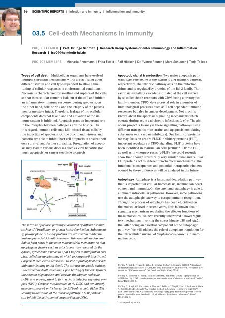
The intrinsic apoptosis pathway is activated by different stimuli<br />
such as UV irradiation or growth factor deprivation. Subsequently,<br />
pro-apoptotic BH3-only proteins are activated to inhibit the<br />
anti-apoptotic Bcl-2 family members. This event allows Bax and<br />
Bak to form pores in the outer mitochondrial membrane so that<br />
apoptogenic factors such as cytochrome c are released. In the<br />
cytosol, cytochrome c binds to Apaf-1 to form a multiprotein complex,<br />
called the apoptosome, at which pro-caspase-9 is activated.<br />
Caspase-9 then cleaves caspase-3 to start a proteolytical cascade<br />
ultimately leading to cell death. The extrinsic apoptosis pathway<br />
is activated by death receptors. Upon binding of trimeric ligands,<br />
the receptor oligomerizes and recruits the adapter molecule<br />
FADD and pro-caspase-8 to form a death inducing signaling complex<br />
(DISC). Caspase-8 is activated at the DISC and can directly<br />
activate caspase-3 or it cleaves the BH3-only protein Bid to tBid<br />
leading to activation of the intrinsic pathway. c-FLIP proteins<br />
can inhibit the activation of caspase-8 at the DISC.<br />
Autophagy Autophagy is a lysosomal degradation pathway<br />
that is important for cellular homeostasis, mammalian development<br />
and immunity. On the one hand, autophagy is able to<br />
eliminate intracellular pathogens. However, some pathogens<br />
use the autophagic pathway to escape immune recognition.<br />
Though the process of autophagy has been elucidated on<br />
the molecular level in recent years, little is known about<br />
signaling mechanisms regulating the effector functions of<br />
these molecules. We have recently uncovered a novel regulatory<br />
mechanism involving the stress kinase p38 and Atg5,<br />
the latter being an essential component of the autophagic<br />
pathway. We will address the role of autophagy regulation for<br />
the intracellular survival of Staphylococcus aureus in mammalian<br />
cells.<br />
Ueffing N, Keil E, Freund C, Kühne R, Schulze-Osthoff K, Schmitz I (2008) “Structural<br />
and mutational analyses of c-FLIPR, the only murine short FLIP isoform, reveal requirements<br />
for DISC recruitment”. Cell Death and Differ 15(4):773-82.<br />
Ueffing N, Schuster M, Keil E, Schulze-Osthoff K, Schmitz I (2008) “Upregulation of<br />
c-FLIPshort by NFAT contributes to apoptosis resistance of short-term activated T cells”.<br />
Blood 112(3):690-8.<br />
Ueffing N, Singh KK, Christians A, Thorns C, Feller AC, Nagl F, Fend F, Heikaus S, Marx<br />
A, Zotz RB, Brade J, Schulz WA, Schulze-Osthoff K, Schmitz I*, Schwerk C (2009) “A<br />
SNP in the cellular FLICE-inhibitory protein (c-FLIP) gene determines protein isoform<br />
production and is associated with risk of follicular lymphoma in humans”. Blood<br />
114(3):572-9.<br />
* corresponding author