

CHAPTER X CHAPTER 4 - Cancer et environnement
CHAPTER X CHAPTER 4 - Cancer et environnement
CHAPTER X CHAPTER 4 - Cancer et environnement

You also want an ePaper? Increase the reach of your titles
YUMPU automatically turns print PDFs into web optimized ePapers that Google loves.
crucial for the development of this cancer,<br />
in particular related to invasive<br />
growth {2236}.<br />
Several candidate genes have been proposed<br />
to explain the gain of 12p in<br />
TGCTs. These included KRAS2, which is<br />
rarely mutated and som<strong>et</strong>imes overexpressed<br />
in TGCTs {1818,1829,1953,<br />
2192,2436}, and cyclin D2 (CCND2)<br />
{1128,2325,2404,2436}. The latter might<br />
be involved via a deregulated G1-S<br />
checkpoint. A more focused approach<br />
to the identification of candidate genes<br />
was initiated by the finding of a m<strong>et</strong>astatic<br />
seminoma with a high level of amplification<br />
of a restricted region of 12p, cytogen<strong>et</strong>ically<br />
identified as 12p11.2-p12.1<br />
{2530}. Subsequently, primary TGCTs<br />
have been found with such an amplification<br />
{1360,1793,1795,2147,2221,2914}.<br />
The 12p-amplicon occurs in about 8-<br />
10% of primary seminomas, particularly<br />
in those lacking an i(12p) {2914}, and it<br />
is much rarer in non-seminomas. This<br />
suggests the existence of two pathways<br />
leading to overrepresentation of certain<br />
genes on 12p, either via isochromosome<br />
formation, or an alternative mechanism,<br />
possibly followed by high level amplification.<br />
The seminomas with amplification<br />
have a reduced sensitivity to apoptosis<br />
for which DAD-R is a promising<br />
candidate {2914}. Probably more genes<br />
on 12p, in particular in the amplicon,<br />
help the tumour cells to overcome apoptosis<br />
{807}.<br />
Molecular gen<strong>et</strong>ic alterations<br />
Multiple studies on the possible role of<br />
inactivation of tumour suppressor genes<br />
and activation of proto-oncogenes in the<br />
development of TGCTs have been<br />
reported. Interpr<strong>et</strong>ation of the findings<br />
must be done with caution if the data<br />
derived from the tumours are compared<br />
to normal testicular parenchyma, which<br />
does not contain the normal counterpart<br />
of the cell of origin of this cancer.<br />
A significant difference in genome m<strong>et</strong>hylation<br />
has been reported b<strong>et</strong>ween seminomas<br />
(hypom<strong>et</strong>hylated) and non-seminomas<br />
(hyperm<strong>et</strong>hylated) {882,2443}.<br />
This could reflect simply their embryonic<br />
origin, and the capacity of the non-seminomas<br />
to mimic embryonal and extra<br />
embryonal development. This is for<br />
example supported by their pattern of<br />
expression of OCT3/4, also known as<br />
POU5F1 {2003} X-inactivation {1538}, as<br />
well as their telomerase activity.<br />
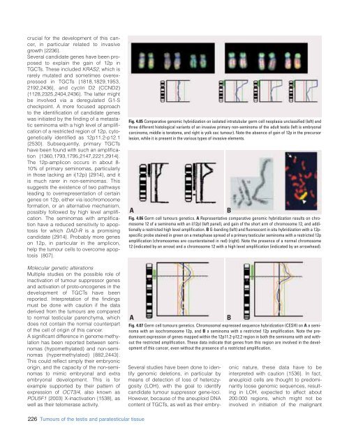
Fig. 4.05 Comparative genomic hybridization on isolated intratubular germ cell neoplasia unclassified (left) and<br />
three different histological variants of an invasive primary non-seminoma of the adult testis (left is embryonal<br />
carcinoma, middle is teratoma, and right is yolk sac tumour). Note the absence of gain of 12p in the precursor<br />
lesion, while it is present in the various types of invasive elements.<br />
A<br />
Fig. 4.06 Germ cell tumours gen<strong>et</strong>ics. A Representative comparative genomic hybridization results on chromosome<br />
12 of a seminoma with an i(12p) (left panel), and gain of the short arm of chromosome 12, and additionally<br />
a restricted high level amplification. B G-banding (left) and fluorescent in situ hybridization with a 12pspecific<br />
probe stained in green on a m<strong>et</strong>aphase spread of a primary testicular seminoma with a restricted 12p<br />
amplification (chromosomes are counterstained in red) (right). Note the presence of a normal chromosome<br />
12 (indicated by an arrow) and a chromosome 12 with a high level amplification (indicated by an arrowhead).<br />
A<br />
Fig. 4.07 Germ cell tumours gen<strong>et</strong>ics. Chromosomal expressed sequence hybridization (CESH) on A a seminoma<br />
with an isochromosome 12p, and B a seminoma with a restricted 12p amplification. Note the predominant<br />
expression of genes mapped within the 12p11.2-p12.2 region in both the seminoma with and without<br />
the restricted amplification. These data indicate that genes from this region are involved in the development<br />
of this cancer, even without the presence of a restricted amplification.<br />
B<br />
B<br />
Several studies have been done to identify<br />
genomic del<strong>et</strong>ions, in particular by<br />
means of d<strong>et</strong>ection of loss of h<strong>et</strong>erozygosity<br />
(LOH), with the goal to identify<br />
candidate tumour suppressor gene-loci.<br />
However, because of the aneuploid DNA<br />
content of TGCTs, as well as their embryonic<br />
nature, these data have to be<br />
interpr<strong>et</strong>ed with caution {1536}. In fact,<br />
aneuploid cells are thought to predominantly<br />
loose genomic sequences, resulting<br />
in LOH, expected to affect about<br />
200.000 regions, which might not be<br />
involved in initiation of the malignant<br />
226 Tumours of the testis and paratesticular tissue


