

CHAPTER X CHAPTER 4 - Cancer et environnement
CHAPTER X CHAPTER 4 - Cancer et environnement
CHAPTER X CHAPTER 4 - Cancer et environnement

Create successful ePaper yourself
Turn your PDF publications into a flip-book with our unique Google optimized e-Paper software.
have cytoplasmic lacunae that contain<br />
pink secr<strong>et</strong>ion or erythrocytes. The<br />
cytotrophoblastic cells have pale to clear<br />
cytoplasm with a single, irregularly<br />
shaped nucleus with one or two prominent<br />
nucleoli. Intermediate trophoblastic<br />
cells have eosinophilic to clear cytoplasm<br />
and single nuclei; they are larger<br />
than cytotrophoblastic cells but may not<br />
be readily discernible from them without<br />
the use of immunohistochemical stains.<br />
A<br />
B<br />
Immunoprofile<br />
The syncytiotrophoblasts are positive for<br />
hCG, alpha subunit of inhibin<br />
{1664,2042} and epithelial membrane<br />
antigen {1894}. Stains for hCG may also<br />
highlight large cells that possibly represent<br />
transitional forms b<strong>et</strong>ween mononucleated<br />
trophoblastic cells and syncytiotrophoblasts.<br />
The intermediate trophoblastic<br />
cells are positive for human<br />
placental lactogen {1615, 1616} and, if<br />
comparable to the gestational examples,<br />
would be expected to stain for Mel-CAM<br />
and HLA-G {2425}. All of the cell types<br />
express cytokeratin, and placental alkaline<br />
phosphatase shows patchy reactivity<br />
in about one half of the cases.<br />
Prognosis<br />
Choriocarcinoma often disseminates<br />
prior to its discovery, probably because<br />
of its propensity to invade blood vessels.<br />
As a consequence, the majority of<br />
patients present with advanced stage<br />
disease. It is this aspect of choriocarcinoma<br />
that causes it to be associated with<br />
a worse prognosis than most other forms<br />
of testicular germ cell tumour. Additionally,<br />
high levels of hCG correlate with a<br />
C<br />
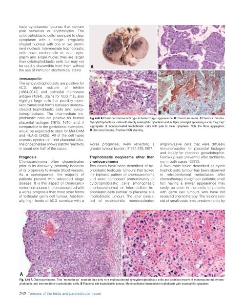
Fig. 4.43 A Choriocarcinoma with typical hemorrhagic appearance. B Choriocarcinoma. C Choriocarcinoma.<br />
Syncytiotrophoblastic cells with deeply eosinophilic cytoplasm and multiple, smudged appearing nuclei; they "cap"<br />
aggregates of mononucleated trophoblastic cells with pale to clear cytoplasm. Note the fibrin aggregates.<br />
D Choriocarcinoma. Positive HCG staining.<br />
worse prognosis, likely reflecting a<br />
greater tumour burden {7,281,575,1897}.<br />
Trophoblastic neoplasms other than<br />
choriocarcinoma<br />
Two cases have been described of trophoblastic<br />
testicular tumours that lacked<br />
the biphasic pattern of choriocarcinoma<br />
and were composed predominantly of<br />
cytotrophoblastic cells (monophasic<br />
choriocarcinoma) or intermediate trophoblastic<br />
cells (similar to placental site<br />
trophoblastic tumour). The latter consisted<br />
of eosinophilic mononucleated<br />
D<br />
angioinvasive cells that were diffusely<br />
immunoreactive for placental lactogen<br />
and focally for chorionic gonadotrophin.<br />
Follow-up was uneventful after orchiectomy<br />
in both cases {2672}.<br />
A favourable lesion described as cystic<br />
trophoblastic tumour has been observed<br />
in r<strong>et</strong>roperitoneal m<strong>et</strong>astases after<br />
chemotherapy in eighteen patients; small<br />
foci having a similar appearance may<br />
rarely be seen in the testis of patients<br />
with germ cell tumours who have not<br />
received chemotherapy. The lesions consist<br />
of small cysts lined predominantly by<br />
A<br />
B<br />
Fig. 4.44 A Choriocarcinoma. This "monophasic" example has only rare multinucleated syncytiotrophoblastic cells and consists mostly of mononucleated cytotrophoblastic<br />
and intermediate trophoblastic cells. B Placental site trophoblastic tumour. Mononucleated intermediate trophoblasts with eosinophilic cytoplasm.<br />
242 Tumours of the testis and paratesticular tissue


