

CHAPTER X CHAPTER 4 - Cancer et environnement
CHAPTER X CHAPTER 4 - Cancer et environnement
CHAPTER X CHAPTER 4 - Cancer et environnement

Create successful ePaper yourself
Turn your PDF publications into a flip-book with our unique Google optimized e-Paper software.
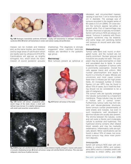
A<br />
Fig. 4.69 Androgen insensitivity syndrome. A Sertoli - Leydig cell hamartomas in androgen insensitivity<br />
syndrome (AIS). B Sertoli cell hamartoma in center and nodular Leydig cell proliferation.<br />
masses can be multiple and bilateral<br />
and, as the name implies, are characterized<br />
by large areas of calcification which<br />
are readily seen by ultrasound {410,873}.<br />
Calcifications will app`ear as brightly<br />
echogenic foci, which block the transmission<br />
of sound (posterior acoustic<br />
Fig. 4.70 Sertoli cell tumour. Longitudinal ultrasound<br />
image of the testis shows a small well<br />
defined mass (arrow). It is slightly h<strong>et</strong>erogeneous<br />
with small cysts (anechoic areas) within it.<br />
A<br />
B<br />
shadowing). This diagnosis is strongly<br />
suggested when calcified testicular<br />
masses are identified in the pediatric<br />
age group.<br />
Macroscopy<br />
Most tumours present as spherical or<br />
Fig. 4.71 Sertoli cell tumour of the testis.<br />
Fig. 4.72 A Large cell calcified Sertoli cell tumour shows bilateral, brightly echogenic masses with posterior<br />
acoustic shadowing (arrow). B Sertoli cell tumour. Large cell calcified Sertoli cell tumour. This case was<br />
malignant; note focus of yellow necrosis.<br />
B<br />
lobulated, well circumscribed masses,<br />
varying in size from 1 cm to more than 20<br />
cm in diam<strong>et</strong>er. The average size of<br />
tumours recorded in the largest series of<br />
60 cases is 3.5 cm {2894}. On cross section<br />
the tumours appear tan-yellow or<br />
greyish white. Foci of haemorrhage may<br />
be seen. Necrosis is typically not evident.<br />
Sertoli cell tumours NOS are always unilateral.<br />
Tumours in patients with Peutz-<br />
Jeghers syndrome may be bilateral,<br />
and some large cell calcifying Sertoli<br />
cell tumours on record were also bilateral<br />
{1391}.<br />
Histopathology<br />
Tumour cells have oval, round, or elongated<br />
nuclei, and the nucleoli are not<br />
overtly prominent. Nuclear grooves and<br />
inclusions are usually not seen. The cytoplasm<br />
may be pale eosinophilic or clear<br />
and vacuolated due to lipids. In some<br />
instances the cytoplasm of tumour cells<br />
is prominently eosinophilic. Overall the<br />
cells appear bland and uniform. Mild<br />
nuclear pleomorphism and atypia is<br />
found in a minority of cases. Mitoses are<br />
uncommon and most cases contain<br />
fewer than 5 mitoses per ten high power<br />
fields. An increased number of mitotic<br />
figures (>5 per HPF) may be found in<br />
about 15% of cases, but in itself this finding<br />
should not be considered to be a<br />
sign of malignancy.<br />
The tumour cells are typically arranged<br />
into tubules surrounded by a basement<br />
membrane. These tubules may be solid<br />
or hollow with a central lumen.<br />
Furthermore, tumour cells may form r<strong>et</strong>iform<br />
and tubular-glandular structures.<br />
Some tumours consist predominantly of<br />
solid she<strong>et</strong>s and nodules, but even in<br />
such neoplasms, well developed or<br />
abortive tubules are usually also present.<br />
The stroma b<strong>et</strong>ween the tubules, cords<br />
and cell nests is fibrotic and moderately<br />
cellular to acellular and hyalinized. The<br />
hyalinized stroma contains often dilated<br />
blood vessels and may be markedly<br />
oedematous. Inflammatory cells are typically<br />
absent. Minor calcifications can be<br />
found in about 10% of cases, but occasional<br />
tumours may show more prominent<br />
deposits.<br />
Immunoprofile<br />
Sertoli cell tumours NOS stain with antibodies<br />
to vimentin (90%) and cytokeratins<br />
(80%) and to a variable extent with<br />
antibodies to inhibin (40%), and S100<br />
Sex cord / gonadal stromal tumours 253


