

CHAPTER X CHAPTER 4 - Cancer et environnement
CHAPTER X CHAPTER 4 - Cancer et environnement
CHAPTER X CHAPTER 4 - Cancer et environnement

Create successful ePaper yourself
Turn your PDF publications into a flip-book with our unique Google optimized e-Paper software.
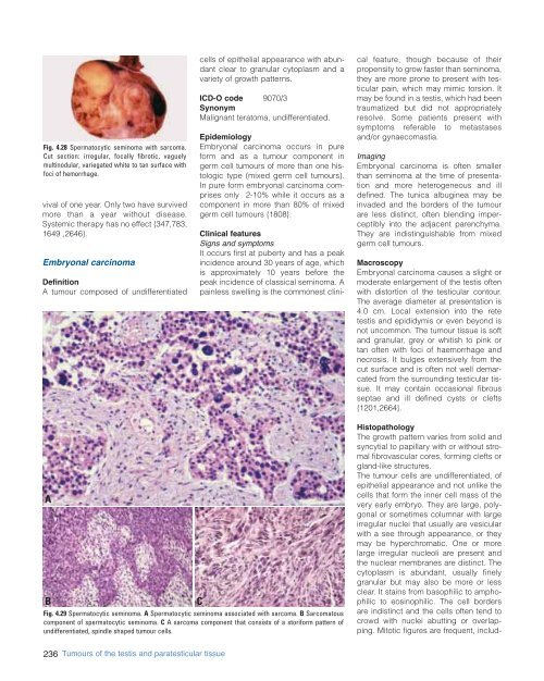
Fig. 4.28 Spermatocytic seminoma with sarcoma.<br />
Cut section: irregular, focally fibrotic, vaguely<br />
multinodular, variegated white to tan surface with<br />
foci of hemorrhage.<br />
vival of one year. Only two have survived<br />
more than a year without disease.<br />
Systemic therapy has no effect {347,783,<br />
1649 ,2646}.<br />
Embryonal carcinoma<br />
Definition<br />
A tumour composed of undifferentiated<br />
cells of epithelial appearance with abundant<br />
clear to granular cytoplasm and a<br />
vari<strong>et</strong>y of growth patterns.<br />
ICD-O code 9070/3<br />
Synonym<br />
Malignant teratoma, undifferentiated.<br />
Epidemiology<br />
Embryonal carcinoma occurs in pure<br />
form and as a tumour component in<br />
germ cell tumours of more than one histologic<br />
type (mixed germ cell tumours).<br />
In pure form embryonal carcinoma comprises<br />
only 2-10% while it occurs as a<br />
component in more than 80% of mixed<br />
germ cell tumours {1808}.<br />
Clinical features<br />
Signs and symptoms<br />
It occurs first at puberty and has a peak<br />
incidence around 30 years of age, which<br />
is approximately 10 years before the<br />
peak incidence of classical seminoma. A<br />
painless swelling is the commonest clinical<br />
feature, though because of their<br />
propensity to grow faster than seminoma,<br />
they are more prone to present with testicular<br />
pain, which may mimic torsion. It<br />
may be found in a testis, which had been<br />
traumatized but did not appropriately<br />
resolve. Some patients present with<br />
symptoms referable to m<strong>et</strong>astases<br />
and/or gynaecomastia.<br />
Imaging<br />
Embryonal carcinoma is often smaller<br />
than seminoma at the time of presentation<br />
and more h<strong>et</strong>erogeneous and ill<br />
defined. The tunica albuginea may be<br />
invaded and the borders of the tumour<br />
are less distinct, often blending imperceptibly<br />
into the adjacent parenchyma.<br />
They are indistinguishable from mixed<br />
germ cell tumours.<br />
Macroscopy<br />
Embryonal carcinoma causes a slight or<br />
moderate enlargement of the testis often<br />
with distortion of the testicular contour.<br />
The average diam<strong>et</strong>er at presentation is<br />
4.0 cm. Local extension into the r<strong>et</strong>e<br />
testis and epididymis or even beyond is<br />
not uncommon. The tumour tissue is soft<br />
and granular, grey or whitish to pink or<br />
tan often with foci of haemorrhage and<br />
necrosis. It bulges extensively from the<br />
cut surface and is often not well demarcated<br />
from the surrounding testicular tissue.<br />
It may contain occasional fibrous<br />
septae and ill defined cysts or clefts<br />
{1201,2664}.<br />
A<br />
B<br />
Fig. 4.29 Spermatocytic seminoma. A Spermatocytic seminoma associated with sarcoma. B Sarcomatous<br />
component of spermatocytic seminoma. C A sarcoma component that consists of a storiform pattern of<br />
undifferentiated, spindle shaped tumour cells.<br />
C<br />
Histopathology<br />
The growth pattern varies from solid and<br />
syncytial to papillary with or without stromal<br />
fibrovascular cores, forming clefts or<br />
gland-like structures.<br />
The tumour cells are undifferentiated, of<br />
epithelial appearance and not unlike the<br />
cells that form the inner cell mass of the<br />
very early embryo. They are large, polygonal<br />
or som<strong>et</strong>imes columnar with large<br />
irregular nuclei that usually are vesicular<br />
with a see through appearance, or they<br />
may be hyperchromatic. One or more<br />
large irregular nucleoli are present and<br />
the nuclear membranes are distinct. The<br />
cytoplasm is abundant, usually finely<br />
granular but may also be more or less<br />
clear. It stains from basophilic to amphophilic<br />
to eosinophilic. The cell borders<br />
are indistinct and the cells often tend to<br />
crowd with nuclei abutting or overlapping.<br />
Mitotic figures are frequent, includ-<br />
236 Tumours of the testis and paratesticular tissue


