

CHAPTER X CHAPTER 4 - Cancer et environnement
CHAPTER X CHAPTER 4 - Cancer et environnement
CHAPTER X CHAPTER 4 - Cancer et environnement

You also want an ePaper? Increase the reach of your titles
YUMPU automatically turns print PDFs into web optimized ePapers that Google loves.
Sex cord / gonadal stromal tumours<br />
I.A. Sesterhenn<br />
J. Cheville<br />
P.J. Woodward<br />
I. Damjanov<br />
G.K. Jacobsen<br />
M. Nistal<br />
R. Paniagua<br />
A.A. Renshaw<br />
Sex cord / gonadal stromal<br />
tumours, pure forms<br />
Included in this category are Leydig cell<br />
tumours, Sertoli cell tumours, granulosa<br />
cell tumours and tumours of the thecoma/fibroma<br />
group.<br />
These tumours constitute about 4-6% of<br />
adult testicular tumours and over 30% of<br />
testicular tumours in infants and children.<br />
The name given to this group does not<br />
indicate a preference for any particular<br />
concept of testicular embryogenesis. As<br />
with the germ cell tumours, the aim<br />
throughout this section is to closely parallel<br />
the WHO terminology and classification<br />
of ovarian tumours.<br />
About 10% of these tumours, almost<br />
always in adults, m<strong>et</strong>astasize. However, it<br />
may not be possible on histological<br />
grounds to forecast their behaviour. Some<br />
of these tumours occur in androgen insensitivity<br />
syndrome (AIS) and adrenogenital<br />
syndrome (AGS) and should be classified<br />
under tumour-like lesions.<br />
Leydig cell tumour<br />
Definition<br />
A tumour composed of elements recapitulating<br />
normal development and evolution<br />
of Leydig cells.<br />
ICD-O codes<br />
Leydig cell tumour 8650/1<br />
Malignant Leydig cell tumour 8650/3<br />
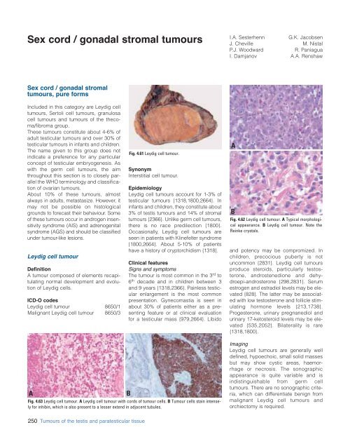
Fig. 4.61 Leydig cell tumour.<br />
Synonym<br />
Interstitial cell tumour.<br />
Epidemiology<br />
Leydig cell tumours account for 1-3% of<br />
testicular tumours {1318,1800,2664}. In<br />
infants and children, they constitute about<br />
3% of testis tumours and 14% of stromal<br />
tumours {2366}. Unlike germ cell tumours,<br />
there is no race predilection {1800}.<br />
Occasionally, Leydig cell tumours are<br />
seen in patients with Klinefelter syndrome<br />
{1800,2664}. About 5-10% of patients<br />
have a history of cryptorchidism {1318}.<br />
Clinical features<br />
Signs and symptoms<br />
The tumour is most common in the 3 rd to<br />
6 th decade and in children b<strong>et</strong>ween 3<br />
and 9 years {1318,2366}. Painless testicular<br />
enlargement is the most common<br />
presentation. Gynecomastia is seen in<br />
about 30% of patients either as a presenting<br />
feature or at clinical evaluation<br />
for a testicular mass {979,2664}. Libido<br />
A<br />
B<br />
Fig. 4.62 Leydig cell tumour. A Typical morphological<br />
appearance. B Leydig cell tumour. Note the<br />
Reinke crystals.<br />
and potency may be compromized. In<br />
children, precocious puberty is not<br />
uncommon {2831}. Leydig cell tumours<br />
produce steroids, particularly testosterone,<br />
androstenedione and dehydroepi-androsterone<br />
{298,2831}. Serum<br />
estrogen and estradiol levels may be elevated<br />
{828}. The latter may be associated<br />
with low testosterone and follicle stimulating<br />
hormone levels {213,1738}.<br />
Progesterone, urinary pregnanediol and<br />
urinary 17-k<strong>et</strong>osteroid levels may be elevated<br />
{535,2052}. Bilaterality is rare<br />
{1318,1800}.<br />
A<br />
Fig. 4.63 Leydig cell tumour. A Leydig cell tumour with cords of tumour cells. B Tumour cells stain intensely<br />
for inhibin, which is also present to a lesser extend in adjacent tubules.<br />
B<br />
Imaging<br />
Leydig cell tumours are generally well<br />
defined, hypoechoic, small solid masses<br />
but may show cystic areas, haemorrhage<br />
or necrosis. The sonographic<br />
appearance is quite variable and is<br />
indistinguishable from germ cell<br />
tumours. There are no sonographic criteria,<br />
which can differentiate benign from<br />
malignant Leydig cell tumours and<br />
orchiectomy is required.<br />
250 Tumours of the testis and paratesticular tissue


