

CHAPTER X CHAPTER 4 - Cancer et environnement
CHAPTER X CHAPTER 4 - Cancer et environnement
CHAPTER X CHAPTER 4 - Cancer et environnement

Create successful ePaper yourself
Turn your PDF publications into a flip-book with our unique Google optimized e-Paper software.
Histologic patterns<br />
Microcystic or r<strong>et</strong>icular pattern<br />
The microcystic pattern consists of a<br />
meshwork of vacuolated cells producing<br />
a honeycomb appearance. The tumour<br />
cells are small and may be compressed<br />
by the vacuoles, which may contain pale<br />
eosinophilic secr<strong>et</strong>ion. The nuclei vary in<br />
size, but generally are small. Mitotic<br />
activity is typically brisk. Hyaline globules<br />
are often present {1201,2593}. This<br />
pattern is the most common one.<br />
Macrocystic pattern<br />
The macrocystic pattern consists of collections<br />
of thin-walled spaces of varying<br />
sizes. They may be adjacent to each other,<br />
or separated by other histologic patterns.<br />
Solid pattern<br />
The solid pattern consists of nodular collections<br />
or aggregates of medium sized<br />
polygonal tumour cells with clear cytoplasm,<br />
prominent nuclei, and usually<br />
showing brisk mitotic activity. It is often<br />
associated with a peripheral microcystic<br />
pattern which helps distinguish it from<br />
typical seminoma and embryonal carcinoma.<br />
Som<strong>et</strong>imes the cells may show<br />
greater pleomorphism and giant cells<br />
may be present.<br />
Glandular-alveolar pattern<br />
This pattern consists of collections of irregular<br />
alveoli, gland-like spaces and tubular<br />
structures lined by cells varying from flattened<br />
to cuboidal or polygonal. The glandlike<br />
spaces or clefts form a meshwork of<br />
cavities and channels, som<strong>et</strong>imes interspersed<br />
with myxomatous tissue.<br />
Endodermal sinus pattern<br />
This pattern consists of structures composed<br />
of a stalk of connective tissue containing<br />
a thin walled blood vessel and<br />
lined on the surface by a layer of cuboidal<br />
cells with clear cytoplasm and prominent<br />
nuclei. Mitotic activity is usually present<br />
and may be brisk. These structures, also<br />
known as Schiller-Duval bodies, are considered<br />
a hallmark of YST {2593}. They<br />
are seen scattered within the tumour in<br />
varying numbers. Their absence does not<br />
preclude the diagnosis.<br />
Papillary pattern<br />
This pattern has numerous, usually fine<br />
papillae consisting of connective tissue<br />
cores lined by cells with prominent nuclei<br />
and often showing brisk mitotic activity.<br />
The connective tissue cores vary from<br />
loose and oedematous to fibrous and<br />
hyalinized. Som<strong>et</strong>imes there may be considerable<br />
deposits of hyaline material<br />
forming wider and more solid brightly<br />
eosinophilic and amorphous papillae.<br />
Myxomatous pattern<br />
This pattern consists of collections of<br />
myxomatous tissue containing sparse<br />
cords, strands or collections of individual<br />
cells showing prominent nuclei and<br />
mitotic activity {1201}.<br />
Polyvesicular vitelline pattern<br />
This pattern consists of collections of<br />
vesicles or cysts varying in shape and<br />
size surrounded by connective tissue<br />
which may vary from cellular and oedematous<br />
to dense and fibrous. The vesicles<br />
are lined by columnar to flattened<br />
cells. Som<strong>et</strong>imes the vesicles are small<br />
and adhere to each other, and some may<br />
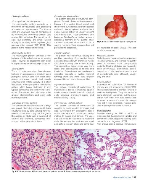
Fig. 4.36 Yolk sac tumour in the testis of a one year old.<br />
be hourglass shaped {2593}. This pattern<br />
is uncommon.<br />
Hepatoid pattern<br />
Collections of hepatoid cells are present<br />
in some tumours, and is more frequently<br />
seen in tumours from postpubertal<br />
patients. Hyaline globules are frequently<br />
seen {1197,2669}. Som<strong>et</strong>imes, collections<br />
of such cells may be numerous and<br />
of considerable size, although usually<br />
they are small.<br />
Enteric pattern<br />
Individual or collections of immature<br />
glands are not uncommon {1201,2669}.<br />
They usually resemble allantois, enteric or<br />
endom<strong>et</strong>rioid glands. They are similar to<br />
some glands in teratomas, but the association<br />
with other yolk sac tumour patterns<br />
and absence of a muscular component<br />
aid in their distinction. Hyaline globules<br />
may be present and numerous.<br />
Immunoprofile<br />
Positive staining for AFP is helpful in<br />
diagnosis but the reaction is variable and<br />
som<strong>et</strong>imes weak. Negative staining does<br />
not exclude a diagnosis of YST.<br />
YST shows strong positive immunocyto-<br />
A<br />
B<br />
Fig. 4.37 Yolk sac tumour. A Microcystic pattern of yolk sac tumour. B Glandular-alveolar pattern of yolk sac tumour.<br />
Germ cell tumours 239


