terminology and guidelines for glaucoma ii - Kwaliteitskoepel
terminology and guidelines for glaucoma ii - Kwaliteitskoepel
terminology and guidelines for glaucoma ii - Kwaliteitskoepel
You also want an ePaper? Increase the reach of your titles
YUMPU automatically turns print PDFs into web optimized ePapers that Google loves.
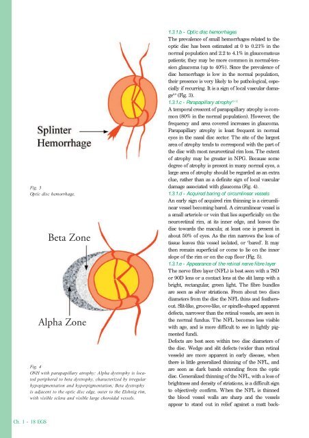
Fig. 3<br />
Optic disc hemorrhage.<br />
Beta Zone<br />
Alpha Zone<br />
Fig. 4<br />
ONH with parapapillary atrophy: Alpha dystrophy is located<br />
peripheral to beta dystrophy, characterized by irregular<br />
hypopigmentation <strong>and</strong> hyperpigmentation; Beta dystrophy<br />
is adjacent to the optic disc edge, outer to the Elshnig rim,<br />
with visible sclera <strong>and</strong> visible large choroidal vessels.<br />
1.3.1.b - Optic disc hemorrhages<br />
The prevalence of small hemorrhages related to the<br />
optic disc has been estimated at 0 to 0.21% in the<br />
normal population <strong>and</strong> 2.2 to 4.1% in <strong>glaucoma</strong>tous<br />
patients; they may be more common in normal-tension<br />
<strong>glaucoma</strong> (up to 40%). Since the prevalence of<br />
disc hemorrhage is low in the normal population,<br />
their presence is very likely to be pathological, especially<br />
if recurring. It is a sign of local vascular damage<br />
8,9 (Fig. 3).<br />
1.3.1.c - Parapapillary atrophy 10-12<br />
A temporal crescent of parapapillary atrophy is common<br />
(80% in the normal population). However, the<br />
frequency <strong>and</strong> area covered increases in <strong>glaucoma</strong>.<br />
Parapapillary atrophy is least frequent in normal<br />
eyes in the nasal disc sector. The site of the largest<br />
area of atrophy tends to correspond with the part of<br />
the disc with most neuroretinal rim loss. The extent<br />
of atrophy may be greater in NPG. Because some<br />
degree of atrophy is present in many normal eyes, a<br />
large area of atrophy should be regarded as an extra<br />
clue, rather than as a definite sign of local vascular<br />
damage associated with <strong>glaucoma</strong> (Fig. 4).<br />
1.3.1.d - Acquired baring of circumlinear vessels<br />
An early sign of acquired rim thinning is a circumlinear<br />
vessel becoming bared. A circumlinear vessel is<br />
a small arteriole or vein that lies superficially on the<br />
neuroretinal rim, at its inner edge, <strong>and</strong> leaves the<br />
disc towards the macula; at least one is present in<br />
about 50% of eyes. As the rim narrows the loss of<br />
tissue leaves this vessel isolated, or ‘bared’. It may<br />
then remain superficial or come to lie on the inner<br />
slope of the rim or on the cup floor (Fig. 5).<br />
1.3.1.e - Appearance of the retinal nerve fibre layer<br />
The nerve fibre layer (NFL) is best seen with a 78D<br />
or 90D lens or a contact lens at the slit lamp with a<br />
bright, rectangular, green light. The fibre bundles<br />
are seen as silver striations. From about two discs<br />
diameters from the disc the NFL thins <strong>and</strong> feathersout.<br />
Slit-like, groove-like, or spindle-shaped apparent<br />
defects, narrower than the retinal vessels, are seen in<br />
the normal fundus. The NFL becomes less visible<br />
with age, <strong>and</strong> is more difficult to see in lightly pigmented<br />
fundi.<br />
Defects are best seen within two disc diameters of<br />
the disc. Wedge <strong>and</strong> slit defects (wider than retinal<br />
vessels) are more apparent in early disease, when<br />
there is little generalized thinning of the NFL, <strong>and</strong><br />
are seen as dark b<strong>and</strong>s extending from the optic<br />
disc. Generalized thinning of the NFL, with a loss of<br />
brightness <strong>and</strong> density of striations, is a difficult sign<br />
to objectively confirm. When the NFL is thinned<br />
the blood vessel walls are sharp <strong>and</strong> the vessels<br />
appear to st<strong>and</strong> out in relief against a matt back-<br />
Ch. 1 - 18 EGS


