Varian Linatron High-Energy X-ray Applications 2007
Varian Linatron High-Energy X-ray Applications 2007
Varian Linatron High-Energy X-ray Applications 2007

You also want an ePaper? Increase the reach of your titles
YUMPU automatically turns print PDFs into web optimized ePapers that Google loves.
When radiographing curved sections, the concave side<br />
should face the source and, when possible, flexible film<br />
holders should be formed to the casting contour. While<br />
this practice tends to improve film/screen contact, it may<br />
increase the image distortion at the edges of the curved<br />
film. To control for this, image quality indicators should<br />
be placed at the extreme widths of the film, and there<br />
should be adequate overlap in adjacent exposures to<br />
ensure complete coverage. While an oblique angle may<br />
be required in some regions, when possible, the X-<strong>ray</strong><br />
beam should enter the casting at right angles to the front<br />
surface, and the film should be set up perpendicular to<br />
the central <strong>ray</strong>.<br />
When the casting thickness varies more than 1/2-inch<br />
(12.7 mm) equivalent steel, the multiple-film technique<br />
can be employed to cope with the varying section<br />
thicknesses. These variations may also create significant<br />
scatter, which can be minimized using special procedures<br />
(see the discussion on scatter control).<br />
Layout and Marking. Radiographic assignments can<br />
involve fairly simple techniques. This can be as simple as<br />
placing the film cassette under or behind the casting and<br />
making the exposure. But, for large, heavy castings such as<br />
turbine bodies, the film placement and radiographic<br />
projections can be complex and may require careful<br />
planning and fixturing.<br />
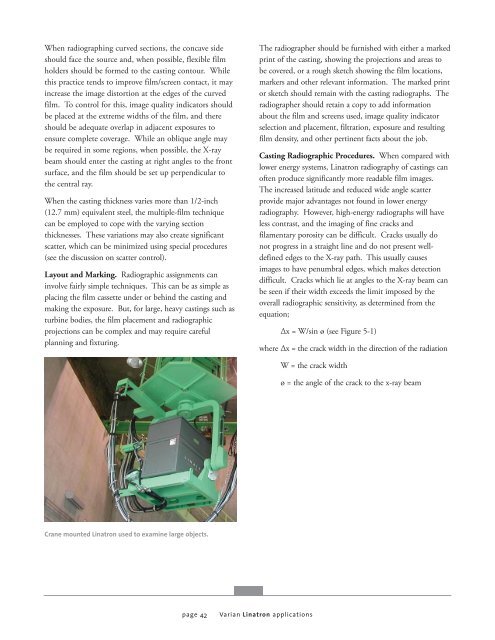
Crane mounted <strong>Linatron</strong> used to examine large objects.<br />
page 42<br />
The radiographer should be furnished with either a marked<br />
print of the casting, showing the projections and areas to<br />
be covered, or a rough sketch showing the film locations,<br />
markers and other relevant information. The marked print<br />
or sketch should remain with the casting radiographs. The<br />
radiographer should retain a copy to add information<br />
about the film and screens used, image quality indicator<br />
selection and placement, filtration, exposure and resulting<br />
film density, and other pertinent facts about the job.<br />
Casting Radiographic Procedures. When compared with<br />
lower energy systems, <strong>Linatron</strong> radiography of castings can<br />
often produce significantly more readable film images.<br />
The increased latitude and reduced wide angle scatter<br />
provide major advantages not found in lower energy<br />
radiography. However, high-energy radiographs will have<br />
less contrast, and the imaging of fine cracks and<br />
filamentary porosity can be difficult. Cracks usually do<br />
not progress in a straight line and do not present welldefined<br />
edges to the X-<strong>ray</strong> path. This usually causes<br />
images to have penumbral edges, which makes detection<br />
difficult. Cracks which lie at angles to the X-<strong>ray</strong> beam can<br />
be seen if their width exceeds the limit imposed by the<br />
overall radiographic sensitivity, as determined from the<br />
equation;<br />
<strong>Varian</strong> <strong>Linatron</strong> applications<br />
Δx = W/sin ø (see Figure 5-1)<br />
where Δx = the crack width in the direction of the radiation<br />
W = the crack width<br />
ø = the angle of the crack to the x-<strong>ray</strong> beam


![[MSDS 126] Dow Corning 200 Fluid, 5 CST Part Number ... - Varian](https://img.yumpu.com/5104917/1/190x245/msds-126-dow-corning-200-fluid-5-cst-part-number-varian.jpg?quality=85)