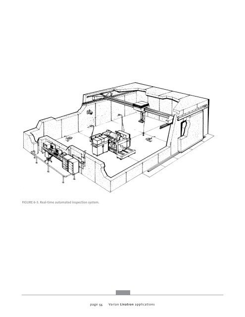
FIGURE 6-3. Real-time automated inspection system. page 56 <strong>Varian</strong> <strong>Linatron</strong> applications
Real-time inspection applications, using proper technique, have been successful for up to twelve half value layers. However, throughput suffers when the long image exposures for this type of imaging are used. Collimators - When real-time imaging is being performed, it is extremely important to restrict the x-<strong>ray</strong> beam to the area that is being inspected. Remote controlled collimators are very useful in real-time radiography because the size and shape of the collimated beam can be seen on the imaging screen in the control room in “real-time”, allowing the operator to select the exact area desired for viewing. Additional collimation may be needed at the object being inspected to control scattered radiation. This can be located either on the x-<strong>ray</strong> side of the object or on the screen side of the object, depending upon the needs of the situation. These are termed “pre-collimators” and “postcollimators”, respectively, and are similar to the practice of “blocking” except that these collimators restrict the beam to the size of the imaging screen, while blocking is only shielding placed around the item being inspected. Manipulators - A key factor in real-time radiography is the ability to manipulate the item under inspection automatically or by remote control. To inspect complicated items, manipulators (or parts handling systems) must be accurately controllable and repeatable. For automated imaging applications, manipulators are programmed to follow predictable, planned paths during the inspection. The design of manipulators is very applications specific and the system integration needed for automated test sequences is challenging. In general, the manipulator must have sufficient flexibility to position the inspected object under inspection properly for the total x-<strong>ray</strong> inspection. Control of the manipulator can be a remote manual console or the controls can be integrated into the imaging system. Integrating manipulator control with the imaging system allows completely autamatic inspection sequences. page 57 Screens/Filters - A distinction should be made between conversion screens used in real-time radiography, which directly convert x-<strong>ray</strong>s to visible light, and intensifying screens, which filter low energy x-<strong>ray</strong>s and emit intensifying photoelectrons to increase light output. Real-time radiography uses several different types of conversion screens. For low energy, these screens can be used without intensifying filters. However, for higher energies, intensifying filters (screens) are used to provide better image contrast. The combination of conversion screen and filter depend upon the application. As higher energies are used, image degradation results from high energy photons passing through the conversion screen without sufficient interaction with the phosphors in the screen. By adding a heavy metal intensifying screen on the x-<strong>ray</strong> side of the fluorescent (conversion) screen, “knock out” electrons are emitted which interact with the phosphors of the screen. These intensifying screens are usually made from tungsten or tantalum. Their thickness requirement is a function of the x-<strong>ray</strong> energy, but in practice 0.020 inch (0.50 mm) thick tantalum is used for energies from 2 to 6 MV and 0.040 inch (1 mm) thick tantalum for higher energies. Fused scintillating fiber optic conversion screens are thicker than conventional fluoroscopic screens and do not generally need intensifying screens. Fiber optic screens exhibit excellent conversion efficiency, which is a function of the screen thickness, and low energy scatter rejection, which is a function of the collimating effect of the individual fibers. Fiber optic screens are especially effective in high scatter, high energy applications. Field of View - Real-time cameras generally provide more than one field of view by using two or more optical lenses to provide magnification. A magnified image results in higher resolution but with a smaller field of view and lower throughput. The application will determine which field of view is appropriate. Image Acquisition - There are generally three basic methods of image acquisition used in real-time radiography, real time, recursive averaging and integration (summation). <strong>Varian</strong> <strong>Linatron</strong> applications


![[MSDS 126] Dow Corning 200 Fluid, 5 CST Part Number ... - Varian](https://img.yumpu.com/5104917/1/190x245/msds-126-dow-corning-200-fluid-5-cst-part-number-varian.jpg?quality=85)