26➔Diagnostic, Patient Monitoring and TherapyComponent Recommendations for ECG/Portable ECG and EEGComponent Recommendations (Continued)Component Description Key Features Benefits Other TI SolutionsProcessors (Continued)MSP430F20xx Ultra Low Power 2 KB Flash, 128 B RAM, SPI+I 2 C 8 ch. 12-bit ADC or 4 ch. 16-bit SD ADC, 4x4 mm16-bit MCUpackageMSP430F22x4 Ultra Low Power 32 KB Flash, 1 KB RAM, SPI + I 2 C + UART/LIN + IrDA 12 ch. 10-bit ADC, 2 op.amps16-bit MCUMSP430F23x0 Ultra Low Power 32 KB Flash, 2 KB RAM, SPI + I 2 C + UART/LIN + IrDA Analog comparator, HW multiplier16-bit MCUMSP430F15x Ultra Low Power 32 KB Flash, 1 KB RAM, SPI + I 2 C + UART, DMA, SVS 8 ch. 12-bit ADC, 2 ch.12-bit DAC, analog comparator16-bit MCUMSP430F16x Ultra Low Power 60 KB Flash, 2 KB RAM, SPI + I 2 C + UART, DMA, SVS 8 ch. 12-bit ADC, 2 ch.12-bit DAC, analog comp,16-bit MCUHW multiplierMSP430F16xx Ultra Low Power 55 KB Flash, 10 KB RAM, SPI + I 2 C + UART, DMA, SVS 8 ch. 12-bit ADC, 2 ch.12-bit DAC, analog comp,16-bit MCUHW multiplierMSP430F41x Ultra Low Power 4 KB Flash, 256 B RAM, SVS, 96 segment LCD Analog comparator16-bit MCUMSP430F42x0 Ultra Low Power 32 KB Flash, 256 B RAM, 56 segment LCD 5 ch. 16-bit SD ADC, 12-bit DAC16-bit MCUMSP430F42x Ultra Low Power 32 KB Flash, 1 KB RAM, SPI + UART, SVS, 128 segment LCD 3 x 16-bit SD ADC16-bit MCUMSP430F43x Ultra Low Power 32 KB Flash, 1 KB RAM, SPI + UART, SVS, 160 segment LCD 8 ch. 12-bit ADC, analog comparator16-bit MCUMSP430FG43x Ultra Low Power 60 KB Flash, 2 KB RAM, SPI + UART, SVS, 128 segment LCD 12 ch. 12-bit ADC, 2 ch. 12-bit DAC, DMA, 3 op amps16-bit MCUMSP430F44x Ultra Low Power 60 KB Flash, 2 KB RAM, 2x SPI + UART, SVS, 160 segment LCD 8 ch. 12-bit ADC, HW multiplier16-bit MCUMSP430FG461x Ultra Low Power 120 KB Flash, 8 KB RAM, SPI + I 2 C + UART/LIN + IrDA, 12 ch.12-bit ADC, 2 ch.12-bit DAC, A-comp, 3 op amp,16-bit MCU 160 LCD HW multiplierPower Management Productsbq20z90 Battery Fuel Gauge Instant accuracy better than 1% error over lifetime of Automatically adjusts for battery aging, battery self bq20z70, bq20z80the batterydischarge and temperature inefficienciesbq24703 Battery Charger 0V operation, ±0.4% charge voltage accuracy, Dynamic power management, multichemistry bq24702integrated PWMbq24721C Battery Charge Multi-chemistry and multi-cell sync switch-mode charger High efficiency, pack and system protection functionsManagementbq29330 Battery Safety Battery pack full-protection analog front end Provides individual cell voltages and battery voltage tobattery management hostDCH010505 Galvanic Isolated, 1W, 3kV isolation, minimal external components Safety isolation, removal of ground loops DCH010512, DCH010515DC/DC ConvertersDCR021205TPS3808 Voltage Supervisor Low quiescent current, programmable-delay Circuit initialization and timing supervision TPS310xTPS54350 DC/DC Converter 4.5 to 20V IN 3A DC/DC with integrated switch FET, Eliminate beat noise/ceramic caps/FPGA/integration TPS54550sync pin, enableTPS65130 Boost Converter 800mA switch, adjustable, dual output, positive Two supplies from one switcherand negative boostTPS79901 Single Channel LDO Very high rejection of power source noise Low-noise power rails for sensitive analog components TPS79501Preview products are listed in bold blue.New products are listed in bold red.<strong>Medical</strong> <strong>Applications</strong> <strong>Guide</strong> Texas Instruments 2Q 2007
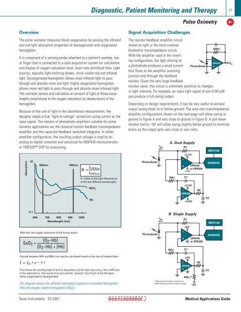
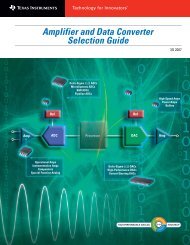
Diagnostic, Patient Monitoring and TherapyPulse Oximetry27➔OverviewThe pulse oximeter measures blood oxygenation by sensing the infraredand red light absorption properties of deoxygenated and oxygenatedhemoglobin.It is comprised of a sensing probe attached to a patient’s earlobe, toeor finger that is connected to a data acquisition system for calculationand display of oxygen saturation level, heart rate and blood flow. Lightsources, typically light-emitting diodes, shine visible red and infraredlight. Deoxygenated hemoglobin allows more infrared light to passthrough and absorbs more red light; highly oxygenated hemoglobinallows more red light to pass through and absorbs more infrared light.The oximeter senses and calculates an amount of light at those wavelengthsproportional to the oxygen saturation (or desaturation) of thehemoglobin.Because of the use of light in the absorbance measurement, thedesigner needs a true “light-to-voltage” conversion using current as theinput signal. The classes of photodiode amplifiers suitable for pulseoximetry applications are the classical resistor-feedback transimpedanceamplifier and the capacitor-feedback switched integrator. In eitheramplifier configuration, the resulting output voltage is read by ananalog-to-digital converter and serialized for MSP430 microcontrolleror TMS320 DSP for processing.Signal Acquisition ChallengesThe resistor-feedback amplifier circuitshown at right is the most commonbioelectric transimpedance circuit.10MΩWith the amplifier used in the invertingconfiguration, the light shining ona photodiode produces a small currentthat flows to the amplifier summingjunction and through the feedbackresistor. Given the very large feedbackPhotodiodeOPA353resistor value, this circuit is extremely sensitive to changesin light intensity. For example, an input light signal of just 0.001µWcan produce a full-swing output.Depending on design requirements, it can be very useful to achieveoutput swing down to or below ground. The auto-zero transimpedanceamplifier configurations shown on the next page will allow swing toground in Figure A and very close to ground in Figure B. A pull-downresistor tied to –5V will allow swing slightly below ground to minimizeerrors as the output gets very close to zero volts.I INA. Dual SupplyR1+2.5VREF3140V OAbsorption10660nm940nmR = I 940nmI 660nmR = Ratio of the Light Absorbanceat the two different wavelengths.HbO 2HbPhotodiode1MΩOPA340or OPA350–2.5VC1+2.5VR2OPA335–2.5VC2ADS83200.1600 700 800 900 1000Wavelength [nm]B. Single SupplyR1+5VREF3140With that, the oxygen saturation of the human blood:SaO 2 =[O 2 –Hb][O 2 –Hb] + [Hb]Photodiode1MΩC1OPA340or OPA350ADS8320(Usually between 94% and 98%) can now be calculated based on the law of Lambert Beer.I = Io • e— ε.xThat shows the resulting light (I) and its dependency of the light source (I o ), the coefficientof the extinction (ε, that results from pic) and the “amount” (χ) of each of the Hb typeseither oxygenated or deoxigenated.I IN–5V+5VOPA33540kΩ*R2C2The diagram shows the different absorption spectra of unloaded hemoglobin(Hb) and oxygen loaded hemoglobin (HbO 2 ).*Optional pull-down resistor toallow below ground output swing.Texas Instruments 2Q 2007www.ti.com/medical<strong>Medical</strong> <strong>Applications</strong> <strong>Guide</strong>
- Page 1 and 2: TMTechnology for InnovatorsMedical
- Page 3 and 4: Consumer and Portable Medical Devic
- Page 6: 6➔Consumer and Portable Medical D
- Page 9 and 10: Consumer and Portable Medical Devic
- Page 11 and 12: Consumer and Portable Medical Devic
- Page 13: Consumer and Portable Medical Devic
- Page 16 and 17: 16➔Consumer and Portable Medical
- Page 18 and 19: 18➔Consumer and Portable Medical
- Page 20 and 21: 20 Diagnostic, Patient Monitoring a
- Page 22 and 23: 22➔Diagnostic, Patient Monitoring
- Page 24 and 25: 24➔Diagnostic, Patient Monitoring
- Page 28 and 29: 28➔Diagnostic, Patient Monitoring
- Page 30 and 31: 30➔Diagnostic, Patient Monitoring
- Page 32 and 33: 32➔Diagnostic, Patient Monitoring
- Page 34 and 35: 34➔Diagnostic, Patient Monitoring
- Page 36 and 37: 36➔Diagnostic, Patient Monitoring
- Page 38 and 39: 38➔Diagnostic, Patient Monitoring
- Page 40 and 41: 40➔Diagnostic, Patient Monitoring
- Page 42 and 43: 42➔Diagnostic, Patient Monitoring
- Page 44 and 45: 44➔Diagnostic, Patient Monitoring
- Page 46 and 47: 46➔Diagnostic, Patient Monitoring
- Page 48 and 49: 48➔Medical ImagingUltrasound/Port
- Page 50 and 51: 50➔Medical ImagingUltrasound/Port
- Page 52 and 53: 52➔Medical ImagingUltrasound/Port
- Page 54 and 55: 54➔Medical ImagingComponent Recom
- Page 56 and 57: 56➔Medical ImagingCT ScannersDual
- Page 58 and 59: 58➔Medical ImagingComponent Recom
- Page 60 and 61: 60➔Medical ImagingMagnetic Resona
- Page 62 and 63: 62➔Medical ImagingMagnetic Resona
- Page 64 and 65: 64➔Medical ImagingComponent Recom
- Page 66 and 67: 66➔Medical ImagingDigital X-Raybl
- Page 68 and 69: 68➔Medical ImagingDigital X-RayLo
- Page 70 and 71: 70➔Medical ImagingPET ScannersPET
- Page 72 and 73: 72➔Medical ImagingComponent Recom
- Page 74 and 75: 74➔Medical ImagingPower Managemen
- Page 76 and 77:
76 Medical Instruments➔Medical In
- Page 78 and 79:
78➔Medical InstrumentsMedical Ins
- Page 80 and 81:
80 Resources➔Enhanced ProductTexa
- Page 82 and 83:
82➔ResourcesTI Design ToolsBelow
- Page 84:
TMTechnology for InnovatorsFindProd