PHYSICS
n - susliks.lv
n - susliks.lv
- No tags were found...

You also want an ePaper? Increase the reach of your titles
YUMPU automatically turns print PDFs into web optimized ePapers that Google loves.
16.4. PROPAGATION OF ACTION POTENTIALS<br />
When a cell is excited and generates an action potential, an<br />
ionic current begin to flow. This process can, in turn, excite<br />
similar neighboring cells. With nerve cells that have a long fiber,<br />
the action potential is generated over a very small segment of the<br />
fiber's length, however, it is propagated in both directions from<br />
the original point of excitation. The rate at which an action potential<br />
moves down a fiber or is propagated from cell to cell is<br />
called the propagation rate. The velocity of the propagation rate<br />
varies widely. The usual velocity range in nerves cells is 20 <br />
140 rn/s; propagation through heart muscle is slower, with an<br />
average rate of 0.2 - 0.4 m/s,<br />
16.5. ELECTROCARDIOGRAPHY<br />
The biopotentials generated by the muscles of the heart result<br />
in the electrocardiogram (abbreviated ECG) and the technique<br />
used to measure these potentials is called electrocardiography.<br />
Each action potential in the heart originates near the top of the<br />
right atrium at a point called the sinoartrial node - a group of<br />
specialized cells that spontaneously generate action potentials at a<br />
regular rate. The action potential propagates in all directions<br />
along the surface of both<br />
atria. A graphic recording<br />
or display of the time-variant<br />
voltages produced by<br />
the myocardium during the<br />
cardiac cycle is called an<br />
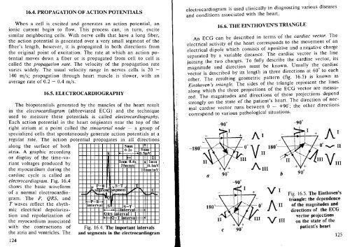
electrocardiogram. Fig. 16.4<br />
shows the basic waveform<br />
of a normal electrocardiogram.<br />
The P, QRS, and<br />
T waves reflect the rhythmic<br />
electrical depolarization<br />
and repolarization of<br />
the myocardium associated<br />
with the contractions of<br />
the atria and ventricles. The<br />
124<br />
R<br />
5mm It I<br />
0.2s f-<br />
emm fo-f0-<br />
0.5mV<br />
1-1 III It<br />
Imm 0.4~ -fij1lmm<br />
25mm/s O.lmV<br />
10mm/mV<br />
P-R S T T<br />
f---P '+---0< ,<br />
....segment segment i.oI" U<br />
-I ~ .I<br />
I I •<br />
fo-P R-<br />
interval S S-T<br />
mterval- -<br />
QRS interval I I<br />
+-t--IO-T interval- -<br />
f--<br />
Fig. 16.4. The important intervals<br />
and segments in the electrocardiogram<br />
z·."<br />
'~'<br />
electrocardiogram is used clinically in diagnosting various diseases<br />
and conditions associated with the heart.<br />
16.6. THE EINTHOVEN'S TRIANGLE<br />
An ECG can be described in terms of the cardiac vector. The<br />
electrical activity of the heart corresponds to the movement of an<br />
electrical di pole which consists of a positive and a negative charge<br />
separated by a variable distance. The cardiac vector is the line<br />
joining the two charges. To fully describe the cardiac vector, its<br />
magnitude and direction must be known. Usually the cardiac<br />
0<br />
vector is described by its length in three directions at 60 to each<br />
other. The resulting geometric pattern (fig. 16.5) is known as<br />
Einthoven 's triangle. The sides of the triangle represent the lines<br />
along which the three projections of the ECG vector are measured.<br />
The magnitudes and directions of these projections depend<br />
strongly on the state of the patient's heart. The direction of normal<br />
cardiac vector runs between 0 - +90'; the other directions<br />
correspond to various pathological situations.<br />
.~ -90 0<br />
~<br />
~'<br />
a<br />
c<br />
AI<br />
AlII<br />
AI<br />
VI Fig. 16.5. The Einthoven's<br />
triangle: the dependence<br />
of the magnitudes and<br />
All directions of the ECG<br />
vector projections<br />
VIII on the state of the<br />
patient's heart<br />
125