PHYSICS
n - susliks.lv
n - susliks.lv
- No tags were found...

Create successful ePaper yourself
Turn your PDF publications into a flip-book with our unique Google optimized e-Paper software.
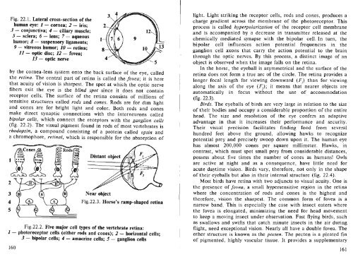
Fig. 22.1. Lateral cross-section of the<br />
human eye: 1 cornea; 2 iris;<br />
3 conjunctiva; 4 ciliary muscle;<br />
5 sclera; 6 lens; 7 aqueous<br />
humor; 8 suspensory ligaments;<br />
9 vitreous humor; 10 retina;<br />
11 optic disc; 12 fovea;<br />
13 optic nerve<br />
by the cornea-lens system onto the back surface of the eye, called<br />
the retina. The central part of retina is called the fovea; it is here<br />
that acuity of vision is sharpest. The spot at which the optic nerve<br />
fibers exit the eye is the blind spot since it does not contain<br />
receptor cells. The surface of the retina consists of millions of<br />
sensitive structures called rods and cones. Rods are for dim light<br />
and cones are for bright light and color. Both rods and cones<br />
make direct synaptic connections with the interneurons called<br />
bipolar cells, which connect the receptors with the ganglion cells<br />
(fig. 22.2). The visual pigment found in rods of most vertebrates is<br />
rhodopsin, a compound consisting of a protein called opsin and<br />
a chromophore, retinal, which is responsible for the absorption of<br />
1<br />
1<br />
Distant object<br />
Near object<br />
Fig.22.3. Horse's ramp-shaped retina<br />
Fig.22.2. Five major cell types of the vertebrate retina:<br />
1 photoreceptor cells (either rods and cones); 2 horizontal cells;<br />
3 - bipolar cells; 4 amacrine cells; 5 ganglion cells<br />
160<br />
light. Light striking the receptor cells, rods and cones, produces a<br />
charge gradient across the membrane of the photoreceptor. This<br />
process is called hyperpolarization of the receptor cell membrane<br />
and is accompanied by a decrease in transmitter released at the<br />
chemically mediated synapse with the bipolar cell. In turn, the<br />
bi polar cell influences action potential frequencies in the<br />
ganglion cell axons that carry the action potential to the brain<br />
through the optic nerves. By this process, a distinct image of an<br />
object is observed when the image falls on the retina.<br />
In the horse, the eyeball is asymmetrical and the surface of the<br />
retina does not form a true arc of the circle. The retina provides a<br />
longer focal length for viewing downward (F) than for viewing<br />
along the axis of the eye (F); it means that nearer objects are<br />
automatically in focus without the use of accommodation<br />
(fig. 22.3).<br />
Birds. The eyeballs of birds are very large in relation to the size<br />
of their bodies and occupy a considerable proportion of the entire<br />
head. The size and resolution of the eye confers an adaptive<br />
advantage in that it increases their performance and security.<br />
Their visual precision facilitates finding food from several<br />
hundred feet above the ground, allowing hawks to recognize<br />
potential prey and precisely swoop down upon it. The human eye<br />
has almost 200,000 cones per square millimeter. Hawks, in<br />
contrast, which must spot small prey from considerable distances,<br />
possess about five times the number of cones as humans! Owls<br />
are active at night and as a consequence, have little need for<br />
acute daytime vision. Birds vary, therefore, not only in the shape<br />
of their eyeballs but also in their internal structure (fig. 22.4).<br />
Most birds have retina with two adjuncts to visual acuity. One is<br />
the presence of fovea, a small hypersensitive region in the retina<br />
where the concentration of rods and cones is the highest and<br />
therefore, vision the sharpest. The common form of fovea is a<br />
narrow band. This is especially the case with insect eaters where<br />
the fovea is elongated, minimizing the need for head movement<br />
to keep a moving insect under observation. Fast flying birds, such<br />
as swallows and swifts that catch minute insects in the air during<br />
flight, need exceptional vision. Nearly all have a double fovea. The<br />
other structure is known as the pecten. The pecten is a pleated fin<br />
of pigmented, highly vascular tissue. It provides a supplementary<br />
161