

Protocols and Applications Guide (US Letter Size) - Promega
Protocols and Applications Guide (US Letter Size) - Promega
Protocols and Applications Guide (US Letter Size) - Promega

Create successful ePaper yourself
Turn your PDF publications into a flip-book with our unique Google optimized e-Paper software.
|| 2RNA Interference<br />
See Figure 2.8, Panels B <strong>and</strong> C for stably transfected cells<br />
that suppressed expression of p53.<br />
B. Delivery of dsRNA to Drosophila S2 Cells in Culture<br />
The protocol outlined below was used to successfully<br />
deliver PCR products of various sizes (180bp or 505bp)<br />
generated either from the 778bp ERK-A target or from a<br />
control plasmid containing the Renilla luciferase gene<br />
(phRL-null Vector; 500bp or 1,000bp) to Drosophila S2 cells<br />
in culture (Figure 2.9; Betz <strong>and</strong> Worzella, 2003). Purified,<br />
in vitro-synthesized ERK-A dsRNA was introduced into<br />
Drosophila S2 cells using the method described by Clemens<br />
et al. (2000) following the protocol described below.<br />
1. Incubate 1 x 10⁶ S2 cells in 1ml of Drosophila expression<br />
system (DES) serum-free medium (Invitrogen) in<br />
triplicate wells of a six-well culture dish in the presence<br />
or absence of various amounts (0, 9.5, 38, or 190nM) of<br />
the test (ERK-A) dsRNA or a nonspecific (Renilla<br />
luciferase) dsRNA.<br />
2. Incubate the S2 cells at room temperature with the<br />
dsRNA for 1 hour, then add 2ml of complete growth<br />
medium.<br />
3. Incubate the cells at room temperature for an additional<br />
3 days to allow for turnover of the target protein.<br />
B<strong>and</strong> Intensity (RFU)<br />
2,500<br />
2,000<br />
1,500<br />
1,000<br />
500<br />
0<br />
180bp<br />
Erk-A<br />
0nM<br />
9.5nM<br />
505bp<br />
Erk-A<br />
dsRNA<br />
778bp<br />
Erk-A<br />
38nM<br />
190nM<br />
500bp<br />
Rluc<br />
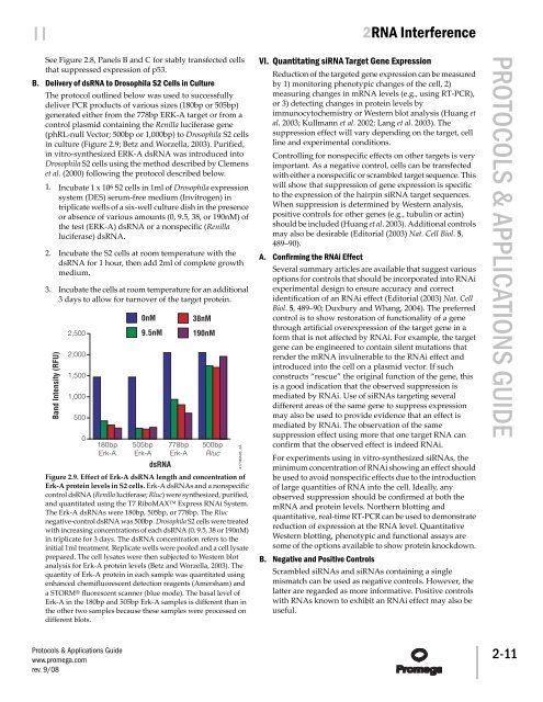
Figure 2.9. Effect of Erk-A dsRNA length <strong>and</strong> concentration of<br />
Erk-A protein levels in S2 cells. Erk-A dsRNAs <strong>and</strong> a nonspecific<br />
control dsRNA (Renilla luciferase; Rluc) were synthesized, purified,<br />
<strong>and</strong> quantitated using the T7 RiboMAX Express RNAi System.<br />
The Erk-A dsRNAs were 180bp, 505bp, or 778bp. The Rluc<br />
negative-control dsRNA was 500bp. Drosophila S2 cells were treated<br />
with increasing concentrations of each dsRNA (0, 9.5, 38 or 190nM)<br />
in triplicate for 3 days. The dsRNA concentration refers to the<br />
initial 1ml treatment. Replicate wells were pooled <strong>and</strong> a cell lysate<br />
prepared. The cell lysates were then subjected to Western blot<br />
analysis for Erk-A protein levels (Betz <strong>and</strong> Worzella, 2003). The<br />
quantity of Erk-A protein in each sample was quantitated using<br />
enhanced chemifluorescent detection reagents (Amersham) <strong>and</strong><br />
a STORM® fluorescent scanner (blue mode). The basal level of<br />
Erk-A in the 180bp <strong>and</strong> 505bp Erk-A samples is different than in<br />
the other two samples because these samples were processed on<br />
different blots.<br />
<strong>Protocols</strong> & <strong>Applications</strong> <strong>Guide</strong><br />
www.promega.com<br />
rev. 9/08<br />
4173MA06_3A<br />
VI. Quantitating siRNA Target Gene Expression<br />
Reduction of the targeted gene expression can be measured<br />
by 1) monitoring phenotypic changes of the cell, 2)<br />
measuring changes in mRNA levels (e.g., using RT-PCR),<br />
or 3) detecting changes in protein levels by<br />
immunocytochemistry or Western blot analysis (Huang et<br />
al. 2003; Kullmann et al. 2002; Lang et al. 2003). The<br />
suppression effect will vary depending on the target, cell<br />
line <strong>and</strong> experimental conditions.<br />
Controlling for nonspecific effects on other targets is very<br />
important. As a negative control, cells can be transfected<br />
with either a nonspecific or scrambled target sequence. This<br />
will show that suppression of gene expression is specific<br />
to the expression of the hairpin siRNA target sequences.<br />
When suppression is determined by Western analysis,<br />
positive controls for other genes (e.g., tubulin or actin)<br />
should be included (Huang et al. 2003). Additional controls<br />
may also be desirable (Editorial (2003) Nat. Cell Biol. 5,<br />
489–90).<br />
A. Confirming the RNAi Effect<br />
Several summary articles are available that suggest various<br />
options for controls that should be incorporated into RNAi<br />
experimental design to ensure accuracy <strong>and</strong> correct<br />
identification of an RNAi effect (Editorial (2003) Nat. Cell<br />
Biol. 5, 489–90; Duxbury <strong>and</strong> Whang, 2004). The preferred<br />
control is to show restoration of functionality of a gene<br />
through artificial overexpression of the target gene in a<br />
form that is not affected by RNAi. For example, the target<br />
gene can be engineered to contain silent mutations that<br />
render the mRNA invulnerable to the RNAi effect <strong>and</strong><br />
introduced into the cell on a plasmid vector. If such<br />
constructs “rescue” the original function of the gene, this<br />
is a good indication that the observed suppression is<br />
mediated by RNAi. Use of siRNAs targeting several<br />
different areas of the same gene to suppress expression<br />
may also be used to provide evidence that an effect is<br />
mediated by RNAi. The observation of the same<br />
suppression effect using more that one target RNA can<br />
confirm that the observed effect is indeed RNAi.<br />
For experiments using in vitro-synthesized siRNAs, the<br />
minimum concentration of RNAi showing an effect should<br />
be used to avoid nonspecific effects due to the introduction<br />
of large quantities of RNA into the cell. Ideally, any<br />
observed suppression should be confirmed at both the<br />
mRNA <strong>and</strong> protein levels. Northern blotting <strong>and</strong><br />
quantitative, real-time RT-PCR can be used to demonstrate<br />
reduction of expression at the RNA level. Quantitative<br />
Western blotting, phenotypic <strong>and</strong> functional assays are<br />
some of the options available to show protein knockdown.<br />
B. Negative <strong>and</strong> Positive Controls<br />
Scrambled siRNAs <strong>and</strong> siRNAs containing a single<br />
mismatch can be used as negative controls. However, the<br />
latter are regarded as more informative. Positive controls<br />
with RNAs known to exhibit an RNAi effect may also be<br />
useful.<br />
PROTOCOLS & APPLICATIONS GUIDE 2-11


