Thesis Title: Subtitle - NMR Spectroscopy Research Group
Thesis Title: Subtitle - NMR Spectroscopy Research Group
Thesis Title: Subtitle - NMR Spectroscopy Research Group

Create successful ePaper yourself
Turn your PDF publications into a flip-book with our unique Google optimized e-Paper software.
3.7 Study case. 87<br />
The coordinates of the A chain in the PDB deposition 2IDO (Kirby et al., 2006) was used as<br />
the structural model for ε186. The structural model of θ was conformer 10 of the <strong>NMR</strong> structure of<br />
θ in complex with ε186 (PDB accession code 2AXD; (Keniry et al., 2006)). This conformer was<br />
chosen because it has the lowest backbone RMSD to the HOT protein (2.1 Å) for residues 9-66 (the<br />
structurally defined region for which meaningful PCS could be measured). The experimentally<br />
determined PCS values of ε186 have been reported previously (Schmitz et al., 2006) and the PCS<br />
values of θ are provided in the Supporting Information. All Δχ-tensor optimizations were performed<br />
using Numbat including the RACS correction term and a tolerance value toli of zero for all spins.<br />
3.7.1 Subunit ε186<br />
Table 3.1 presents the results of the Δχ-tensor fit to the PCS measured for ε186. Initially,<br />
individual eight-variable Δχ-tensor optimizations were performed using the PCS data of each<br />
lanthanide (Table 3.1, columns 1 and 2). Next, the Numbat GUI was updated to display the<br />
deviations between the experimental and back-calculated PCS for the Δχ-tensors found. Several<br />
atoms showed deviations > 0.15 ppm between the experimental and back-calculated PCS (15 out of<br />
199 and 8 out of 255 atoms in the case of Dy 3+ and Er 3+ , respectively. Without the RACS<br />
correction, deviations > 0.15 ppm where observed for 36 and 7 atoms, respectively). Assuming that<br />
these outliers were due to problematic measurements or inaccuracies of the 3D structure, these PCS<br />
were removed interactively using the GUI. Re-calculation of the Δχ-tensor was found not to change<br />
the fitted Δχ-tensor parameters significantly for any of the lanthanide ions (results not shown). This<br />
can be explained by the high quality and large number of experimental PCS data available for each<br />
lanthanide (backbone 13 C’, 15 N and 1 H N spins), resulting in robust fits of the Δχ-tensors.<br />
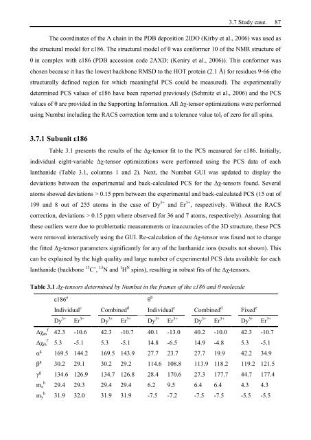
Table 3.1 Δχ-tensors determined by Numbat in the frames of the ε186 and θ molecule<br />
ε186 a θ b<br />
Individual c Combined d Individual c Combined d Fixed e<br />
Dy 3+ Er 3+ Dy 3+ Er 3+ Dy 3+ Er 3+ Dy 3+ Er 3+ Dy 3+ Er 3+<br />
Δχax f 42.3 -10.6 42.3 -10.7 40.1 -13.0 40.2 -10.0 42.3 -10.7<br />
Δχrh f 5.3 -5.1 5.3 -5.1 14.8 -6.5 14.9 -4.8 5.3 -5.1<br />
α g 169.5 144.2 169.5 143.9 27.7 23.7 27.7 19.9 42.2 34.9<br />
β g 30.2 29.1 30.2 29.2 114.6 108.8 113.9 118.2 119.2 121.5<br />
γ g 134.6 126.9 134.7 126.8 28.4 170.6 27.3 177.7 44.7 177.4<br />
mx h 29.4 29.3 29.4 29.4 6.2 9.5 6.4 6.4 4.3 4.3<br />
my h 31.9 32.0 31.9 31.9 -7.5 -7.2 -7.5 -7.5 -5.5 -5.5