Thesis Title: Subtitle - NMR Spectroscopy Research Group
Thesis Title: Subtitle - NMR Spectroscopy Research Group
Thesis Title: Subtitle - NMR Spectroscopy Research Group

Create successful ePaper yourself
Turn your PDF publications into a flip-book with our unique Google optimized e-Paper software.
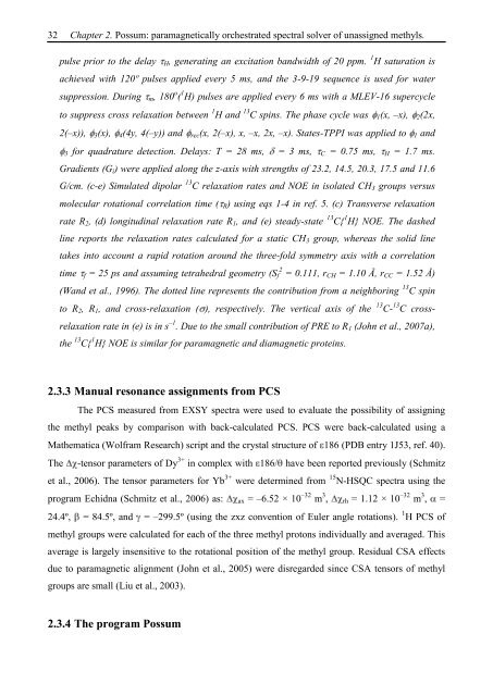
32 Chapter 2. Possum: paramagnetically orchestrated spectral solver of unassigned methyls.<br />
pulse prior to the delay H, generating an excitation bandwidth of 20 ppm. 1 H saturation is<br />
achieved with 120º pulses applied every 5 ms, and the 3-9-19 sequence is used for water<br />
suppression. During m, 180 o ( 1 H) pulses are applied every 6 ms with a MLEV-16 supercycle<br />
to suppress cross relaxation between 1 H and 13 C spins. The phase cycle was 1(x, –x), 2(2x,<br />
2(–x)), 3(x), 4(4y, 4(–y)) and rec(x, 2(–x), x, –x, 2x, –x). States-TPPI was applied to 1 and<br />
3 for quadrature detection. Delays: T = 28 ms, = 3 ms, C = 0.75 ms, H = 1.7 ms.<br />
Gradients (Gi) were applied along the z-axis with strengths of 23.2, 14.5, 20.3, 17.5 and 11.6<br />
G/cm. (c-e) Simulated dipolar 13 C relaxation rates and NOE in isolated CH3 groups versus<br />
molecular rotational correlation time ( R) using eqs 1-4 in ref. 5. (c) Transverse relaxation<br />
rate R2, (d) longitudinal relaxation rate R1, and (e) steady-state 13 C{ 1 H} NOE. The dashed<br />
line reports the relaxation rates calculated for a static CH3 group, whereas the solid line<br />
takes into account a rapid rotation around the three-fold symmetry axis with a correlation<br />
time f = 25 ps and assuming tetrahedral geometry (Sf 2 = 0.111, rCH = 1.10 Å, rCC = 1.52 Å)<br />
(Wand et al., 1996). The dotted line represents the contribution from a neighboring 13 C spin<br />
to R2, R1, and cross-relaxation ( ), respectively. The vertical axis of the 13 C- 13 C cross-<br />
relaxation rate in (e) is in s –1 . Due to the small contribution of PRE to R1 (John et al., 2007a),<br />
the 13 C{ 1 H} NOE is similar for paramagnetic and diamagnetic proteins.<br />
2.3.3 Manual resonance assignments from PCS<br />
The PCS measured from EXSY spectra were used to evaluate the possibility of assigning<br />
the methyl peaks by comparison with back-calculated PCS. PCS were back-calculated using a<br />
Mathematica (Wolfram <strong>Research</strong>) script and the crystal structure of 186 (PDB entry 1J53, ref. 40).<br />
The -tensor parameters of Dy 3+ in complex with 186/ have been reported previously (Schmitz<br />
et al., 2006). The tensor parameters for Yb 3+ were determined from 15 N-HSQC spectra using the<br />
program Echidna (Schmitz et al., 2006) as: ax = –6.52 × 10 –32 m 3 , rh = 1.12 × 10 –32 m 3 , =<br />
24.4º, = 84.5º, and = –299.5º (using the zxz convention of Euler angle rotations). 1 H PCS of<br />
methyl groups were calculated for each of the three methyl protons individually and averaged. This<br />
average is largely insensitive to the rotational position of the methyl group. Residual CSA effects<br />
due to paramagnetic alignment (John et al., 2005) were disregarded since CSA tensors of methyl<br />
groups are small (Liu et al., 2003).<br />
2.3.4 The program Possum