Thesis Title: Subtitle - NMR Spectroscopy Research Group
Thesis Title: Subtitle - NMR Spectroscopy Research Group
Thesis Title: Subtitle - NMR Spectroscopy Research Group
Create successful ePaper yourself
Turn your PDF publications into a flip-book with our unique Google optimized e-Paper software.
xii<br />
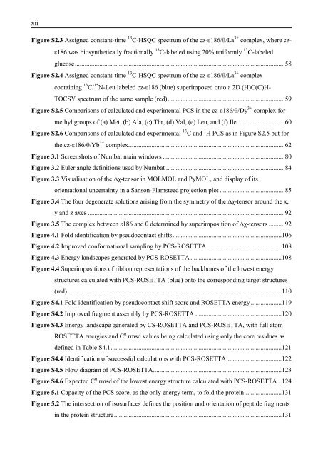
Figure S2.3 Assigned constant-time 13 C-HSQC spectrum of the cz- 186/ /La 3+ complex, where cz-<br />
186 was biosynthetically fractionally 13 C-labeled using 20% uniformly 13 C-labeled<br />
glucose ................................................................................................................................. 58<br />
Figure S2.4 Assigned constant-time 13 C-HSQC spectrum of the cz- 186/ /La 3+ complex<br />
containing 13 C/ 15 N-Leu labeled cz- 186 (blue) superimposed onto a 2D (H)C(C)H-<br />
TOCSY spectrum of the same sample (red) ........................................................................ 59<br />
Figure S2.5 Comparisons of calculated and experimental PCS in the cz- 186/ /Dy 3+ complex for<br />
methyl groups of (a) Met, (b) Ala, (c) Thr, (d) Val, (e) Leu, and (f) Ile ............................. 60<br />
Figure S2.6 Comparisons of calculated and experimental 13 C and 1 H PCS as in Figure S2.5 but for<br />
the cz- 186/ /Yb 3+ complex. ............................................................................................... 62<br />
Figure 3.1 Screenshots of Numbat main windows ........................................................................... 80<br />
Figure 3.2 Euler angle definitions used by Numbat ......................................................................... 84<br />
Figure 3.3 Visualisation of the Δχ-tensor in MOLMOL and PyMOL, and display of its<br />
orientational uncertainty in a Sanson-Flamsteed projection plot ........................................ 85<br />
Figure 3.4 The four degenerate solutions arising from the symmetry of the Δχ-tensor around the x,<br />
y and z axes ......................................................................................................................... 92<br />
Figure 3.5 The complex between ε186 and θ determined by superimposition of Δχ-tensors .......... 92<br />
Figure 4.1 Fold identification by pseudocontact shifts ................................................................... 106<br />
Figure 4.2 Improved conformational sampling by PCS-ROSETTA .............................................. 108<br />
Figure 4.3 Energy landscapes generated by PCS-ROSETTA ........................................................ 108<br />
Figure 4.4 Superimpositions of ribbon representations of the backbones of the lowest energy<br />
structures calculated with PCS-ROSETTA (blue) onto the corresponding target structures<br />
(red) ................................................................................................................................... 110<br />
Figure S4.1 Fold identification by pseudocontact shift score and ROSETTA energy ................... 119<br />
Figure S4.2 Improved fragment assembly by PCS-ROSETTA ..................................................... 120<br />
Figure S4.3 Energy landscape generated by CS-ROSETTA and PCS-ROSETTA, with full atom<br />
ROSETTA energies and C α rmsd values being calculated using only the core residues as<br />
defined in Table S4.1 ......................................................................................................... 121<br />
Figure S4.4 Identification of successful calculations with PCS-ROSETTA .................................. 122<br />
Figure S4.5 Flow diagram of PCS-ROSETTA ............................................................................... 123<br />
Figure S4.6 Expected C α rmsd of the lowest energy structure calculated with PCS-ROSETTA .. 124<br />
Figure 5.1 Capacity of the PCS score, as the only energy term, to fold the protein ....................... 131<br />
Figure 5.2 The intersection of isosurfaces defines the position and orientation of peptide fragments<br />
in the protein structure ....................................................................................................... 131


