Thesis Title: Subtitle - NMR Spectroscopy Research Group
Thesis Title: Subtitle - NMR Spectroscopy Research Group
Thesis Title: Subtitle - NMR Spectroscopy Research Group

You also want an ePaper? Increase the reach of your titles
YUMPU automatically turns print PDFs into web optimized ePapers that Google loves.
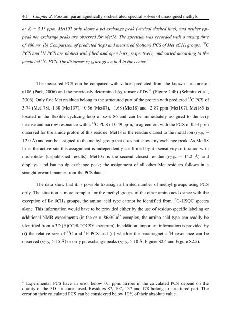
40 Chapter 2. Possum: paramagnetically orchestrated spectral solver of unassigned methyls.<br />
at 2 = 5.53 ppm. Met107 only shows a pd exchange peak (vertical dashed line), and neither pp-<br />
peak nor exchange peaks are observed for Met18. The spectrum was recorded with a mixing time<br />
of 480 ms. (b) Comparison of predicted (top) and measured (bottom) PCS of Met CH3 groups. 13 C<br />
PCS and 1 H PCS are plotted with filled and open bars, respectively, and sorted according to the<br />
predicted 13 C PCS. The distances rC-Ln are given in Å in the center. 3<br />
The measured PCS can be compared with values predicted from the known structure of<br />
186 (Park, 2006) and the previously determined tensor of Dy 3+ (Figure 2.4b) (Schmitz et al.,<br />
2006). Only five Met residues belong to the structured part of the protein with predicted 13 C PCS of<br />
3.74 (Met178), 1.30 (Met137), –0.56 (Met87), –1.68 (Met18) and –2.87 ppm (Met107). Met185 is<br />
located in the flexible cyclizing loop of cz- 186 and can be immediately assigned to the very<br />
intense and narrow resonance with a 13 C PCS of 0.49 ppm, in agreement with the PCS of 0.53 ppm<br />
observed for the amide proton of this residue. Met18 is the residue closest to the metal ion (rC-Dy =<br />
12.0 Å) and can be assigned to the methyl group that does not show any exchange peak. As Met18<br />
lines the active site this assignment is independently confirmed by its sensitivity to titration with<br />
nucleotides (unpublished results). Met107 is the second closest residue (rC-Dy = 14.2 Å) and<br />
displays a pd but no dp exchange peak; the assignment of all other Met residues follows in a<br />
straightforward manner from the PCS data.<br />
The data show that it is possible to assign a limited number of methyl groups using PCS<br />
only. The situation is more complex for the methyl groups of the other amino acids since with the<br />
exception of Ile CH3 groups, the amino acid type cannot be identified from 13 C-HSQC spectra<br />
alone. This information would have to be provided either by the use of residue-specific labeling or<br />
additional <strong>NMR</strong> experiments (in the cz- 186/ /La 3+ complex, the amino acid type can readily be<br />
identified from a 3D (H)CCH-TOCSY spectrum). In addition, important information is provided by<br />
(i) the relative size of 13 C and 1 H PCS and (ii) whether the paramagnetic 1 H resonance can be<br />
observed (rC-Dy > 15 Å) or only pd exchange peaks (rC-Dy > 10 Å, Figure S2.4 and Figure S2.5).<br />
3 Experimental PCS have an error below 0.1 ppm. Errors in the calculated PCS depend on the<br />
quality of the 3D structures used. Residues 87, 107, 137 and 178 belong to structured part. The<br />
error on their calculated PCS can be considered below 10% of their absolute value.