Thesis Title: Subtitle - NMR Spectroscopy Research Group
Thesis Title: Subtitle - NMR Spectroscopy Research Group
Thesis Title: Subtitle - NMR Spectroscopy Research Group
You also want an ePaper? Increase the reach of your titles
YUMPU automatically turns print PDFs into web optimized ePapers that Google loves.
4.2 Introduction. 103<br />
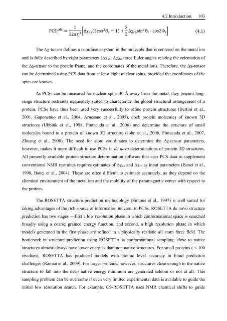
(4.1)<br />
The -tensor defines a coordinate system in the molecule that is centered on the metal ion<br />
and is fully described by eight parameters ( ax, rh, three Euler angles relating the orientation of<br />
the Δχ-tensor to the protein frame, and the coordinates of the metal ion). Therefore, the Δχ-tensor<br />
can be determined using PCS data from at least eight nuclear spins, provided the coordinates of the<br />
spins are known.<br />
As PCSs can be measured for nuclear spins 40 Å away from the metal, they present long-<br />
range structure restraints exquisitely suited to characterize the global structural arrangement of a<br />
protein. PCSs have thus been used very successfully to refine protein structures (Bertini et al.,<br />
2001, Gaponenko et al., 2004, Arnesano et al., 2005), dock protein molecules of known 3D<br />
structures (Ubbink et al., 1998, Pintacuda et al., 2006) and determine the structure of small<br />
molecules bound to a protein of known 3D structure (John et al., 2006, Pintacuda et al., 2007,<br />
Zhuang et al., 2008). The need for atom coordinates to determine the Δχ-tensor parameters,<br />
however, makes it more difficult to use PCSs in de novo determinations of protein 3D structures.<br />
All presently available protein structure determination software that uses PCS data to supplement<br />
conventional <strong>NMR</strong> restraints requires estimates of ax and rh as input parameters (Banci et al.,<br />
1998, Banci et al., 2004). These are often difficult to estimate accurately, as they depend on the<br />
chemical environment of the metal ion and the mobility of the paramagnetic center with respect to<br />
the protein.<br />
The ROSETTA structure prediction methodology (Simons et al., 1997) is well suited for<br />
taking advantages of the rich source of information inherent in PCSs. ROSETTA de novo structure<br />
prediction has two stages —first a low resolution phase in which conformational space is searched<br />
broadly using a coarse grained energy function, and second, a high resolution phase in which<br />
models generated in the first phase are refined in a physically realistic all atom force field. The<br />
bottleneck in structure prediction using ROSETTA is conformational sampling; close to native<br />
structures almost always have lower energies than non native structures. For small proteins ( < 100<br />
residues), ROSETTA has produced models with atomic level accuracy in blind prediction<br />
challenges (Raman et al., 2009). For larger proteins, however, structures close enough to the native<br />
structure to fall into the deep native energy minimum are generated seldom or not at all. This<br />
sampling problem can be overcome if even very limited experimental data is available to guide the<br />
initial low resolution search. For example, CS-ROSETTA uses <strong>NMR</strong> chemical shifts to guide


