- Page 2:
Forensic Pathology for Police, Deat
- Page 5 and 6:
Joseph Prahlow Indiana University S
- Page 7 and 8:
vi Dedication accepted the free gif
- Page 9 and 10:
viii Foreword quality photographs i
- Page 11 and 12:
x Acknowledgements Counties, as wel
- Page 14 and 15:
Contents Part I Introductory Topics
- Page 16 and 17:
Contents xv Hepatobiliary System .
- Page 18 and 19:
Contents xvii RespiratorySystem....
- Page 20 and 21:
Contents xix Mercury ..............
- Page 22 and 23:
Contents xxi Strangulation . ......
- Page 24 and 25:
Contents xxiii Negligence/Neglect .
- Page 26:
About the Author Joseph A. Prahlow,
- Page 29 and 30:
Chapter 1 Introduction to Pathology
- Page 31 and 32:
Introduction to Pathology 5 Table 1
- Page 33 and 34:
Introduction to Pathology 7 Fig. 1.
- Page 35 and 36:
Introduction to Pathology 9 Fig. 1.
- Page 37 and 38:
Introduction to Pathology 11 Fig. 1
- Page 39 and 40:
Introduction to Pathology 13 Fig. 1
- Page 41:
Selected References 15 Disc Image L
- Page 44 and 45:
18 2 Introduction to Forensic Scien
- Page 46 and 47:
20 2 Introduction to Forensic Scien
- Page 48 and 49:
22 2 Introduction to Forensic Scien
- Page 50 and 51:
24 2 Introduction to Forensic Scien
- Page 52 and 53:
26 2 Introduction to Forensic Scien
- Page 54 and 55:
28 2 Introduction to Forensic Scien
- Page 56 and 57:
30 2 Introduction to Forensic Scien
- Page 58 and 59:
32 2 Introduction to Forensic Scien
- Page 61 and 62:
Chapter 3 Introduction to Forensic
- Page 63 and 64:
Duties of the Forensic Pathologist
- Page 65 and 66:
Duties of the Forensic Pathologist
- Page 67 and 68:
Duties of the Forensic Pathologist
- Page 69 and 70:
Training and Qualifications of Fore
- Page 71 and 72:
Training and Qualifications of Fore
- Page 73:
Selected References 47 Selected Ref
- Page 76 and 77:
50 4 Death Investigation Death Inve
- Page 78 and 79:
52 4 Death Investigation met in ord
- Page 80 and 81:
54 4 Death Investigation leading to
- Page 82 and 83:
56 4 Death Investigation Fig. 4.2 A
- Page 84 and 85:
58 4 Death Investigation Fig. 4.5 P
- Page 86 and 87:
60 4 Death Investigation Fig. 4.9 I
- Page 88 and 89:
62 4 Death Investigation apartment
- Page 90 and 91:
64 5 Death Certification Fig. 5.1 A
- Page 92 and 93:
66 5 Death Certification Fig. 5.4 A
- Page 94 and 95:
68 5 Death Certification latter, th
- Page 96 and 97:
70 5 Death Certification infrequent
- Page 98 and 99:
72 5 Death Certification changes in
- Page 100 and 101:
74 5 Death Certification suicides.
- Page 102 and 103:
76 5 Death Certification death is i
- Page 104 and 105:
78 5 Death Certification use involv
- Page 107 and 108:
Chapter 6 Overview of Anatomy and P
- Page 109 and 110:
Introduction 83 Table 6.1 (continue
- Page 111 and 112:
Introduction 85 Table 6.1 (continue
- Page 113 and 114:
Introduction 87 Table 6.1 (continue
- Page 115 and 116:
Introduction 89 Term Definition Tab
- Page 117 and 118:
Introduction 91 “mucous membranes
- Page 119 and 120:
Specific Organ Systems 93 functions
- Page 121 and 122:
Specific Organ Systems 95 Fig. 6.2
- Page 123 and 124:
Specific Organ Systems 97 Microscop
- Page 125 and 126:
Specific Organ Systems 99 Fig. 6.7
- Page 127 and 128:
Specific Organ Systems 101 Fig. 6.1
- Page 129 and 130:
Specific Organ Systems 103 Fig. 6.1
- Page 131 and 132:
Specific Organ Systems 105 Fig. 6.1
- Page 133 and 134:
Specific Organ Systems 107 respirat
- Page 135 and 136:
Specific Organ Systems 109 Fig. 6.1
- Page 137 and 138:
Specific Organ Systems 111 water co
- Page 139 and 140:
Specific Organ Systems 113 Fig. 6.2
- Page 141 and 142:
Specific Organ Systems 115 Platelet
- Page 143 and 144:
Specific Organ Systems 117 Fig. 6.2
- Page 145 and 146:
Specific Organ Systems 119 Fig. 6.2
- Page 147 and 148:
Specific Organ Systems 121 Fig. 6.3
- Page 149 and 150:
Specific Organ Systems 123 Fig. 6.3
- Page 151 and 152:
Disc Image Legends 125 Disc Image 6
- Page 154 and 155:
Part II General Topics in Forensic
- Page 156 and 157:
130 7 The Postmortem Forensic Exami
- Page 158 and 159:
132 7 The Postmortem Forensic Exami
- Page 160 and 161:
134 7 The Postmortem Forensic Exami
- Page 162 and 163:
136 7 The Postmortem Forensic Exami
- Page 164 and 165:
138 7 The Postmortem Forensic Exami
- Page 166 and 167:
140 7 The Postmortem Forensic Exami
- Page 168 and 169:
142 7 The Postmortem Forensic Exami
- Page 170 and 171:
144 7 The Postmortem Forensic Exami
- Page 172 and 173:
146 7 The Postmortem Forensic Exami
- Page 174 and 175:
148 7 The Postmortem Forensic Exami
- Page 176 and 177:
150 7 The Postmortem Forensic Exami
- Page 178 and 179:
152 7 The Postmortem Forensic Exami
- Page 180 and 181:
154 7 The Postmortem Forensic Exami
- Page 182 and 183:
156 7 The Postmortem Forensic Exami
- Page 184 and 185:
158 7 The Postmortem Forensic Exami
- Page 186 and 187:
160 7 The Postmortem Forensic Exami
- Page 189 and 190:
Chapter 8 Postmortem Changes and Ti
- Page 191 and 192:
Early Postmortem Changes 165 Fig. 8
- Page 193 and 194: Early Postmortem Changes 167 can be
- Page 195 and 196: Decomposition 169 exposed to sunlig
- Page 197 and 198: Decomposition 171 Fig. 8.8 As decom
- Page 199 and 200: Decomposition 173 Fig. 8.12 As deco
- Page 201 and 202: Decomposition 175 Fig. 8.16 If flie
- Page 203 and 204: Postmortem Injuries 177 Fig. 8.20 I
- Page 205 and 206: Time of Death Estimation 179 Time o
- Page 207 and 208: Time of Death Estimation 181 Fig. 8
- Page 209 and 210: Disc Image Legends 183 Disc Image 8
- Page 211 and 212: Chapter 9 Identification of Human R
- Page 213 and 214: Introduction 187 Fig. 9.2 Badly dec
- Page 215 and 216: Common, Non-scientific Methods of I
- Page 217 and 218: Scientific Methods of Identificatio
- Page 219 and 220: Scientific Methods of Identificatio
- Page 221 and 222: Identification Based on Other Uniqu
- Page 223 and 224: Identification Based on Other Uniqu
- Page 225 and 226: Unidentified Remains 199 Fig. 9.16
- Page 227: Selected References 201 Disc Image
- Page 231 and 232: Chapter 10 Natural Deaths One man d
- Page 233 and 234: Infectious Disease 207 to as “hyp
- Page 235 and 236: Cardiovascular System 209 refers to
- Page 237 and 238: Cardiovascular System 211 related t
- Page 239 and 240: Cardiovascular System 213 heart int
- Page 241 and 242: Cardiovascular System 215 Aortic An
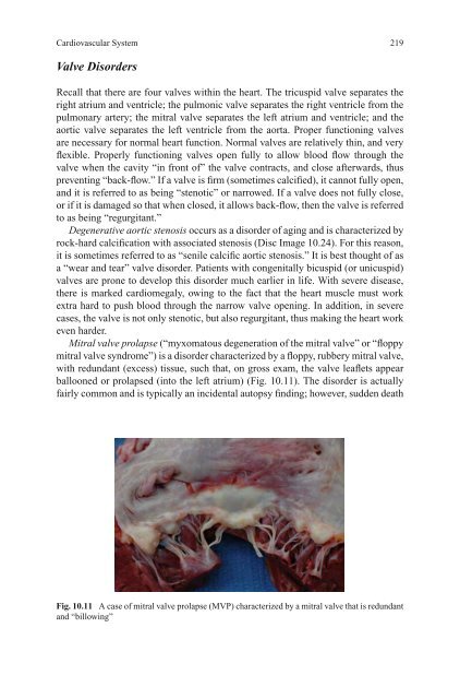
- Page 243: Cardiovascular System 217 Vasculiti
- Page 247 and 248: Cardiovascular System 221 Fig. 10.1
- Page 249 and 250: Central Nervous System 223 Neoplast
- Page 251 and 252: Central Nervous System 225 Viral en
- Page 253 and 254: Central Nervous System 227 Spontane
- Page 255 and 256: Respiratory System 229 Fig. 10.18 T
- Page 257 and 258: Respiratory System 231 Pulmonary Th
- Page 259 and 260: Respiratory System 233 identificati
- Page 261 and 262: Respiratory System 235 Chronic Lung
- Page 263 and 264: Gastrointestinal and Hepatobiliary
- Page 265 and 266: Gastrointestinal and Hepatobiliary
- Page 267 and 268: Reticuloendothelial and Immune Syst
- Page 269 and 270: Endocrine System 243 infection, dis
- Page 271 and 272: Multisystem and Other Disorders 245
- Page 273 and 274: Multisystem and Other Disorders 247
- Page 275 and 276: Multisystem and Other Disorders 249
- Page 277 and 278: Multisystem and Other Disorders 251
- Page 279 and 280: Disc Image Legends 253 Disc Image 1
- Page 281: Selected References 255 Fineschi V,
- Page 284 and 285: 258 11 Drug-Related and Toxin-Relat
- Page 286 and 287: 260 11 Drug-Related and Toxin-Relat
- Page 288 and 289: 262 11 Drug-Related and Toxin-Relat
- Page 290 and 291: 264 11 Drug-Related and Toxin-Relat
- Page 292 and 293: 266 11 Drug-Related and Toxin-Relat
- Page 294 and 295:
268 11 Drug-Related and Toxin-Relat
- Page 296 and 297:
270 11 Drug-Related and Toxin-Relat
- Page 298 and 299:
272 11 Drug-Related and Toxin-Relat
- Page 300 and 301:
274 11 Drug-Related and Toxin-Relat
- Page 302 and 303:
276 11 Drug-Related and Toxin-Relat
- Page 304 and 305:
278 11 Drug-Related and Toxin-Relat
- Page 306 and 307:
280 11 Drug-Related and Toxin-Relat
- Page 308 and 309:
282 11 Drug-Related and Toxin-Relat
- Page 310 and 311:
284 11 Drug-Related and Toxin-Relat
- Page 312 and 313:
286 11 Drug-Related and Toxin-Relat
- Page 314 and 315:
288 11 Drug-Related and Toxin-Relat
- Page 316 and 317:
290 11 Drug-Related and Toxin-Relat
- Page 318 and 319:
292 11 Drug-Related and Toxin-Relat
- Page 320 and 321:
294 11 Drug-Related and Toxin-Relat
- Page 322 and 323:
296 11 Drug-Related and Toxin-Relat
- Page 324 and 325:
298 11 Drug-Related and Toxin-Relat
- Page 327 and 328:
Chapter 12 Blunt Force Injury Death
- Page 329 and 330:
Classification of Blunt Force Injur
- Page 331 and 332:
Classification of Blunt Force Injur
- Page 333 and 334:
Classification of Blunt Force Injur
- Page 335 and 336:
Classification of Blunt Force Injur
- Page 337 and 338:
Blunt Force Head and Neck Trauma 31
- Page 339 and 340:
Blunt Force Head and Neck Trauma 31
- Page 341 and 342:
Blunt Force Head and Neck Trauma 31
- Page 343 and 344:
Blunt Force Head and Neck Trauma 31
- Page 345 and 346:
Blunt Force Head and Neck Trauma 31
- Page 347 and 348:
Blunt Force Head and Neck Trauma 32
- Page 349 and 350:
Special Topics Related to Blunt For
- Page 351 and 352:
Special Topics Related to Blunt For
- Page 353 and 354:
Special Topics Related to Blunt For
- Page 355 and 356:
Special Topics Related to Blunt For
- Page 357 and 358:
Special Topics Related to Blunt For
- Page 359 and 360:
Disc Image Legends 333 Disc Image 1
- Page 361:
Selected References 335 Toro K, Szl
- Page 364 and 365:
338 13 Gunshot Wound Deaths shotgun
- Page 366 and 367:
340 13 Gunshot Wound Deaths Fig. 13
- Page 368 and 369:
342 13 Gunshot Wound Deaths Fig. 13
- Page 370 and 371:
344 13 Gunshot Wound Deaths Fig. 13
- Page 372 and 373:
346 13 Gunshot Wound Deaths targets
- Page 374 and 375:
348 13 Gunshot Wound Deaths (a) (b)
- Page 376 and 377:
350 13 Gunshot Wound Deaths Image 1
- Page 378 and 379:
352 13 Gunshot Wound Deaths Fig. 13
- Page 380 and 381:
354 13 Gunshot Wound Deaths Miscell
- Page 382 and 383:
356 13 Gunshot Wound Deaths Fig. 13
- Page 384 and 385:
358 13 Gunshot Wound Deaths Fig. 13
- Page 386 and 387:
360 13 Gunshot Wound Deaths Fig. 13
- Page 388 and 389:
362 13 Gunshot Wound Deaths may be
- Page 390 and 391:
364 13 Gunshot Wound Deaths Fig. 13
- Page 392 and 393:
366 13 Gunshot Wound Deaths Fig. 13
- Page 394 and 395:
368 13 Gunshot Wound Deaths X-Rays
- Page 396 and 397:
370 13 Gunshot Wound Deaths left-ha
- Page 398 and 399:
372 13 Gunshot Wound Deaths NYCLAD
- Page 400 and 401:
374 13 Gunshot Wound Deaths Disc Im
- Page 402 and 403:
376 13 Gunshot Wound Deaths Disc Im
- Page 405 and 406:
Chapter 14 Sharp Force Injury Death
- Page 407 and 408:
Stab Wounds 381 to describe the sha
- Page 409 and 410:
Stab Wounds 383 describing “clust
- Page 411 and 412:
Incised Wounds 385 Fig. 14.9 A supe
- Page 413 and 414:
Incised Wounds 387 Although knives
- Page 415 and 416:
Special Issues 389 Fig. 14.16 A sta
- Page 417 and 418:
Special Issues 391 Defensive Wounds
- Page 419 and 420:
Special Issues 393 disorders; howev
- Page 421 and 422:
Special Issues 395 area of “radio
- Page 423 and 424:
Special Issues 397 Artifacts A fina
- Page 425 and 426:
Selected References 399 Disc Image
- Page 427 and 428:
Chapter 15 Asphyxial Deaths So Juda
- Page 429 and 430:
Suffocation 403 Fig. 15.1 Petechiae
- Page 431 and 432:
Suffocation 405 Certification of th
- Page 433 and 434:
Suffocation 407 usual occluding sub
- Page 435 and 436:
Suffocation 409 being able to expan
- Page 437 and 438:
Neck Compression (Strangulation) 41
- Page 439 and 440:
Neck Compression (Strangulation) 41
- Page 441 and 442:
Neck Compression (Strangulation) 41
- Page 443 and 444:
Neck Compression (Strangulation) 41
- Page 445 and 446:
Chemical Asphyxia 419 the object ha
- Page 447 and 448:
Chemical Asphyxia 421 Fig. 15.21 A
- Page 449 and 450:
Other Issues 423 to function proper
- Page 451 and 452:
Other Issues 425 (a) (b) Fig. 15.24
- Page 453 and 454:
Other Issues 427 the arms/hands bou
- Page 455 and 456:
Disc Image Legends 429 Disc Image 1
- Page 457:
Selected References 431 Prahlow JA,
- Page 460 and 461:
434 16 Drowning Physiology and Mech
- Page 462 and 463:
436 16 Drowning Fig. 16.2 Thedriver
- Page 464 and 465:
438 16 Drowning Fig. 16.4 “Cadave
- Page 466 and 467:
440 16 Drowning Finding water withi
- Page 468 and 469:
442 16 Drowning Fig. 16.10 Marked d
- Page 470 and 471:
444 16 Drowning Fig. 16.14 True ant
- Page 472 and 473:
446 16 Drowning Manner of Death A l
- Page 474 and 475:
448 16 Drowning Pachar JV, Cameron
- Page 476 and 477:
450 17 Electrical Deaths Fig. 17.1
- Page 478 and 479:
452 17 Electrical Deaths required f
- Page 480 and 481:
454 17 Electrical Deaths death (Fig
- Page 482 and 483:
456 17 Electrical Deaths Fig. 17.8
- Page 484 and 485:
458 17 Electrical Deaths Fig. 17.12
- Page 486 and 487:
460 17 Electrical Deaths Fig. 17.16
- Page 488 and 489:
462 17 Electrical Deaths Fig. 17.20
- Page 490 and 491:
464 17 Electrical Deaths Fig. 17.24
- Page 492 and 493:
466 17 Electrical Deaths Disc Image
- Page 495 and 496:
Chapter 18 Temperature-Related Deat
- Page 497 and 498:
Hypothermia 471 constrict, thus con
- Page 499 and 500:
Hypothermia 473 Fig. 18.3 Prominent
- Page 501 and 502:
Hyperthermia 475 Toxicology testing
- Page 503 and 504:
Hyperthermia 477 Fig. 18.8 A 24-yea
- Page 505 and 506:
Selected References 479 disease. In
- Page 507 and 508:
Chapter 19 Burns and Fire-Related D
- Page 509 and 510:
Introduction 483 Fig. 19.3 Example
- Page 511 and 512:
Burn Types 485 Fig. 19.5 A postmort
- Page 513 and 514:
Burn Types 487 Chemical Burns As me
- Page 515 and 516:
Fire Deaths 489 Fire burns represen
- Page 517 and 518:
Fire Deaths 491 Fig. 19.11 Dense so
- Page 519 and 520:
Fire Deaths 493 Fig. 19.13 Multiple
- Page 521 and 522:
Fire Deaths 495 a CO test or an ins
- Page 523 and 524:
Fire Deaths 497 Fig. 19.20 Atrue an
- Page 525 and 526:
Disc Image Legends 499 Disc Image L
- Page 527 and 528:
Chapter 20 Deaths in Infancy and Ch
- Page 529 and 530:
Discarded Fetuses/Infants and Fetal
- Page 531 and 532:
Discarded Fetuses/Infants and Fetal
- Page 533 and 534:
Birth-Related Infant Deaths 507 abr
- Page 535 and 536:
Infant Deaths 509 Fig. 20.3 A diaph
- Page 537 and 538:
Infant Deaths 511 Fig. 20.6 The pre
- Page 539 and 540:
Natural Death in Childhood 513 path
- Page 541 and 542:
Accidental Childhood Deaths 515 pon
- Page 543 and 544:
Homicidal Childhood Deaths 517 occu
- Page 545 and 546:
Homicidal Childhood Deaths 519 Fig.
- Page 547 and 548:
Homicidal Childhood Deaths 521 in a
- Page 549 and 550:
Homicidal Childhood Deaths 523 Fig.
- Page 551 and 552:
Homicidal Childhood Deaths 525 Fig.
- Page 553 and 554:
Classification of Childhood Homicid
- Page 555 and 556:
Classification of Childhood Homicid
- Page 557 and 558:
Classification of Childhood Homicid
- Page 559 and 560:
Pediatric Autopsy Considerations 53
- Page 561 and 562:
Disc Image Legends 535 Disc Image L
- Page 563:
Selected References 537 Selected Re
- Page 566 and 567:
540 21 Miscellaneous Topics Aircraf
- Page 568 and 569:
542 21 Miscellaneous Topics CAMI to
- Page 570 and 571:
544 21 Miscellaneous Topics To summ
- Page 572 and 573:
546 21 Miscellaneous Topics typical
- Page 574 and 575:
548 21 Miscellaneous Topics quite o
- Page 576 and 577:
550 21 Miscellaneous Topics Fig. 21
- Page 578 and 579:
552 21 Miscellaneous Topics The emb
- Page 580 and 581:
554 21 Miscellaneous Topics various
- Page 582 and 583:
556 21 Miscellaneous Topics stress
- Page 584 and 585:
558 21 Miscellaneous Topics Fig. 21
- Page 586 and 587:
560 21 Miscellaneous Topics ampheta
- Page 588 and 589:
562 21 Miscellaneous Topics victim
- Page 590 and 591:
564 21 Miscellaneous Topics more se
- Page 592 and 593:
566 21 Miscellaneous Topics Fig. 21
- Page 594 and 595:
568 21 Miscellaneous Topics Organ a
- Page 596 and 597:
570 21 Miscellaneous Topics Fig. 21
- Page 598 and 599:
572 21 Miscellaneous Topics region
- Page 600 and 601:
574 21 Miscellaneous Topics Regardi
- Page 602 and 603:
576 21 Miscellaneous Topics and lar
- Page 604 and 605:
578 21 Miscellaneous Topics some wo
- Page 606 and 607:
580 21 Miscellaneous Topics Non-ion
- Page 608 and 609:
582 21 Miscellaneous Topics Yersini
- Page 610 and 611:
584 21 Miscellaneous Topics water (
- Page 612 and 613:
586 21 Miscellaneous Topics to gene
- Page 614 and 615:
588 21 Miscellaneous Topics Disc Im
- Page 616 and 617:
590 21 Miscellaneous Topics Leclair
- Page 619 and 620:
Appendix: Additional Resources and
- Page 621 and 622:
Index Note: The letters ‘f’ and
- Page 623 and 624:
Index 597 Arnold-Chiari malformatio
- Page 625 and 626:
Index 599 closed head injuries, 311
- Page 627 and 628:
Index 601 immediate, 68 intermediar
- Page 629 and 630:
Index 603 liver demonstrating cirrh
- Page 631 and 632:
Index 605 Death investigation syste
- Page 633 and 634:
Index 607 pressure related processe
- Page 635 and 636:
Index 609 bridging veins, 316 dura
- Page 637 and 638:
Index 611 brain, serially-sectioned
- Page 639 and 640:
Index 613 Gestational DM, 247 Gesta
- Page 641 and 642:
Index 615 Homicide(s), 444 by heart
- Page 643 and 644:
Index 617 Infant death, 501-502, 50
- Page 645 and 646:
Index 619 Material Safety Data Shee
- Page 647 and 648:
Index 621 CNS, 223-229 cerebral pal
- Page 649 and 650:
Index 623 Over-the-counter (OTC), 2
- Page 651 and 652:
Index 625 Pregnancy-related materna
- Page 653 and 654:
Index 627 industrial settings, 486
- Page 655 and 656:
Index 629 Smoke and soot inhalation
- Page 657 and 658:
Index 631 Thrombophlebitis, inflamm