- Page 1: ^r»i^/ -•-JP, i>J> > > > 'jr J.
- Page 7 and 8: J i(( THE ) JOURNAL OF BOTANY BEITI
- Page 9 and 10: C. C. Babington, M.A., F.R.S. J. E.
- Page 12 and 13: sydnev Parkinson dd. 1 .Tri-umfetta
- Page 14 and 15: 2 EPILOBIUM NOTES FOR 1880. Cocos I
- Page 16 and 17: 4 EPILOBIUM NOTES FOR 1889. allegat
- Page 18 and 19: 6 EPILOBIUM NOTES FOK 1880. It seem
- Page 20 and 21: 8 EPILOBIUM NOTES FOR 1889. within
- Page 22 and 23: 10 MARINE ALG.E OF THE ARBROATH DIS
- Page 24 and 25: 12 MARINE ALO.E OF THE ARBROATH DIS
- Page 26 and 27: 14 MARINE ALG.E OF THE ARBROATH DIS
- Page 28 and 29: 16 SYNOPSIS OF GENERA AND SPECIES O
- Page 30 and 31: 18 CYPERUS JEMINICUS ROTTB. An annu
- Page 32 and 33: 20 BIOGRAPHICAL INDEX OF BRITISH AN
- Page 34 and 35: 22 SHORT NOTES. 16G0. Master of Mec
- Page 36 and 37: 24 HANDBOOK OF THE BROMELIACEi^. pu
- Page 38 and 39: 26 HANDBOOK OF TIfE BROMELIACE^. wh
- Page 40 and 41: 28 NAMES AND SYNONYMS OF BRITISH PL
- Page 42 and 43: 30 MANUAL OF ORCHIDACEOUS PLANTS. i
- Page 44: 32 LINNEAN SOCIETY OF LONDON. Oeste
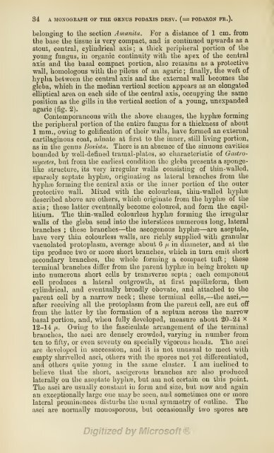
- Page 48 and 49: Gr.Massee del. R.Morgajilith. Podau
- Page 52 and 53: 86 A MONOGRAPH OF THE GENUS PODAXIS
- Page 54 and 55: 38 A MONOGRAPH OP THE GENUS PODAXIS
- Page 56 and 57: 40 NOTES ON SCOTCH PLANTS. prevente
- Page 58 and 59: 42 NOTES ON SCOTCH PLANTS. umhrosa
- Page 60 and 61: 44 NOTES ON SCOTCH PLANTS. Tofieldi
- Page 62 and 63: 46 NOTES ON SCOTCH PLANTS, a sheep-
- Page 64 and 65: 48 ON FKSTUOA HETEROPHYLLA. quality
- Page 66 and 67: 50 ON FESTUCA HETEEOPHYLLA. (( Char
- Page 68 and 69: 52 BIOGRAPHICAL INDEX OF BRITISH AN
- Page 70 and 71: 54 BIOGRAPHICAL, INDEX OP BRITISH A
- Page 72 and 73: 56 BIOGRAMICAL INDEX OF BRITISH AND
- Page 74 and 75: t^ La Bl'oLOGIE V^GETALE. lucidity
- Page 76 and 77: 60 ATLAS DEUTSOHER MEERESALGEN. do
- Page 78 and 79: 62 ATLAS DEUTSCHKR MEERESALGEN. of
- Page 80: 64 LINNEAN SOCIETY OF LONDON. vulga
- Page 83 and 84: 05 NOTES ON THE BRITISH CHARACEJE F
- Page 85 and 86: XOTES ON TIIK BRITISH CHARACE.T: FO
- Page 87 and 88: A MONOGRAPH OF THE GENUS PODAXIS DE
- Page 89 and 90: A MONOGRAPH OF THE GENUS PODAXIS DE
- Page 91 and 92: A JIOXOGEAPH OF THE GENUS PODAXIS D
- Page 93 and 94: A MONOGRAPH OF THE GENUS PODAXIS DE
- Page 95 and 96: A MONOGRAPH OF THE GENUS PODAXIS DE
- Page 97 and 98: FURTHER RECORDS FROM ICELAND. 79 Ba
- Page 99 and 100: FURTHER RECORDS FROM ICELAND. 81 dn
- Page 101 and 102:
PUETHER BECOBDS FROM ICELAND. 88 JE
- Page 103 and 104:
BENJAMIN CLAEKE, F.L.S. 85 (lateral
- Page 105 and 106:
PLANTS FOUND NEAK KILMANOCK, CO. WE
- Page 107 and 108:
BIOGRAPHICAL INDEX OF BRITISH AND I
- Page 109 and 110:
SHORT NOTES. 91 Univ., Dublin, 1845
- Page 111 and 112:
FLORA OF SUFFOLK. 93 hence notice o
- Page 113 and 114:
AKTICLES IN JOURNALS. 95 des Bering
- Page 115 and 116:
97 NOTES ON ENGLISH RUBI. By W. 0.
- Page 117 and 118:
NOTES OX ENGLISH RUBI. 99 The only
- Page 119 and 120:
NOTES ON' ENGLISH RUBI. 101 ones ar
- Page 121 and 122:
VASCULAR CRYPTOGAMIA 01' NEW GUINEA
- Page 123 and 124:
VASCULAR CRYPTOGAMIA OF NEW GUINEA.
- Page 125 and 126:
VASCULAK CRYPTOGAMIA OF NKW GUINEA.
- Page 127 and 128:
VASCULAR CRYPTOGAMIA OF NEW GUINEA.
- Page 129 and 130:
PLANTS 1-UI:ND IN KEIIRY. Ill Jitnc
- Page 131 and 132:
PLANTS FOUND IN KERRY. 118 C. praco
- Page 133 and 134:
PLANTS FOUND IN KERRY. 115 Reeks ;
- Page 135 and 136:
BIOGEAPHICAL INDEX OF BRITISH AND I
- Page 137 and 138:
BIOGEAPHICAL INDEX OF BRITISH AND I
- Page 139 and 140:
SCIENTIFIC PAPERS OF ASA GRAY. 121
- Page 141 and 142:
ARTICLES IN JOURNALS; 123 Wilson's
- Page 143 and 144:
LIKNEAN SOCIETY OF LONDON. 125 perf
- Page 145 and 146:
LINNEAX SOCIETY OF LONDON. 127 spac
- Page 147 and 148:
129 NOTES ON ENGLISH RUBI. By W. 0.
- Page 149 and 150:
NOTES ON ENGLISH RUBI. 131 the livi
- Page 151 and 152:
NOTES ON EN(iUSH KUHI. 133 8G. R. M
- Page 153 and 154:
THE GENUS SCAPHOSEPALUM PFITZER. 13
- Page 155 and 156:
NOTKS ON PONDWEEDS. 137 G. S. PULVi
- Page 157 and 158:
NOTES ON PONDWEEDS. 139 A specimen
- Page 159 and 160:
SYNOPSIS OF OENRUA AST) SPKCIKS OF
- Page 161 and 162:
SYNOPSIS OF GENERA AND SPECIES OF M
- Page 163 and 164:
A NEW LASTREA FROM ASSAM. 145 petal
- Page 165 and 166:
MARINE ALG^ OF DEVON. 147 have now
- Page 167 and 168:
149 THE GENEEA OF STAPELIE^. The mo
- Page 169 and 170:
BIOGRAPHICAL INDEX OF BRITISH AND I
- Page 171 and 172:
BiOGEAPHICAL INDEX OF BRITISH AND I
- Page 173 and 174:
BIOGRAPHICAL INDEX OF BRITISH AND I
- Page 175 and 176:
157 SHOKT NOTES. Lejeunea Rossettia
- Page 177 and 178:
ARTICLES IN JOURNALS, 159 White Smu
- Page 180 and 181:
R.Morgan lith. X 8 ^^^'- l-S-Cantha
- Page 182 and 183:
162 NOTES ON A NEW SUBSPECIES OF EU
- Page 184 and 185:
164 NOTES ON A NEW SUBSPECIES OF EU
- Page 186 and 187:
166 SHORT DESCRIPTIVE NOTES ON THRE
- Page 188 and 189:
168 ABERDEEN, tORFAR, AND DUMFRIES
- Page 190 and 191:
170 ABERDEEN, FOEFAE, AND DUMFRIES
- Page 192 and 193:
172 CAREX RIGIDA AND ITS VARIETIES.
- Page 194 and 195:
174 SUPPOSED HYBEIDITY IN POTAMOGET
- Page 196 and 197:
176 SUPPOSED HYBRIDITY IN POTAMOGET
- Page 198 and 199:
178 SUPPOSED HYBRIDITY IN POTAMOGET
- Page 200 and 201:
180 NOTES ON HIGHLAND PLANTS. conne
- Page 202 and 203:
182 NOTES ON mGHLAND PLANTS. 1886,
- Page 204 and 205:
i84 BIOGRAPHICAL INDEX OF BRITISH A
- Page 206 and 207:
186 BIOGRAPHICAL INDEX OF BRITISH A
- Page 208 and 209:
188 sdoE5: Nol^s. Robinsor, James F
- Page 210 and 211:
190 NOTICES OF BOOKS. Physiology. A
- Page 212 and 213:
192 LINNEAN SOCIETY OF LONDON. Flor
- Page 214 and 215:
194 SYNOPSIS OF THE GENUS TUNICA. c
- Page 216 and 217:
196 ' SYNOPSIS OF THE GENUS TUNICA.
- Page 218 and 219:
198 SYNOPSIS OF THE GENUS TUNICA. p
- Page 220 and 221:
200 HEPATIC.E FOUND IN KERRY, 1889.
- Page 222 and 223:
202 HEPATIC.E FOUND IN KERRY, 1889.
- Page 224 and 225:
204 EUBUS EEYTHRINUS. It may be wel
- Page 226 and 227:
206 KUBUS ERYTIIRINUS. There are fe
- Page 228 and 229:
208 SYNOPSIS OF GENERA AND SPECIES
- Page 230 and 231:
210 SYNOPSIS OF GENERA AND SPECIES
- Page 232 and 233:
212 SYNOPSIS OF GENERA AND SPECIES
- Page 234 and 235:
214 PUCCINIA DIGIlAPUll)iS. cismal
- Page 236 and 237:
216 CHANGES AT KEW. upper cell thic
- Page 238 and 239:
2l8 SHORT NOTES. were clustered a l
- Page 240 and 241:
220 NOTICES OF nooKs. following opi
- Page 242 and 243:
222 LINNEAN SOCIETY OF LONDON, nov.
- Page 244 and 245:
224 LINNEAN SOCIETY OF LONDON. tion
- Page 246 and 247:
226 NOTES ON PONDWEEDS, occupied sp
- Page 248 and 249:
228 NOTES ON OXFORD PLANTS. *Fumana
- Page 250 and 251:
230 NOTES ON OXFORD PLANTS. f. apri
- Page 252 and 253:
232 NOTES ON OXFORD PLANTS. Bor. No
- Page 254 and 255:
234 ON SPAUGANIUM. Bhclmum spicant
- Page 256 and 257:
23G ON SPARGANIUM. perhaps, not alt
- Page 258 and 259:
238 ADDITIONS TO THE IRISH MOSS FLO
- Page 260 and 261:
240 SYNOPSIS OF GENERA AND SPECIES
- Page 262 and 263:
242 SYNOPSIS OF GENERA AND SPECIES
- Page 264 and 265:
244 BIOGRAPHICAL INDEX OF RRITISH A
- Page 266 and 267:
21G BIOGRAPHICAL INDEX. OF BRITISH
- Page 268 and 269:
218 SHOUT NOTES. 447. Herbaiium and
- Page 270 and 271:
250 NOTICES OF BOOKS. Introduction
- Page 272 and 273:
252 NOTICES OF BOOKS. author's prev
- Page 274 and 275:
254 NOTICES OF BOOKS. be grateful t
- Page 276:
256 LINNEAN SOCIETY OF LONDON. e lo
- Page 279 and 280:
257 ON SOME RUSTS AND MILDEWS IX IN
- Page 281 and 282:
ON SOME BUSTS AND MILDEWS IN INDIA.
- Page 283 and 284:
ON SOME RUSTS AND MILDEWS IN INDIA.
- Page 285 and 286:
TONQUIN FERNS. 263 li-2 ft. loDg, 4
- Page 287 and 288:
TONQUIN FERNS. 2G5 broad. Veins pin
- Page 289 and 290:
36, 100, 101, 1872. G. elliptica Ba
- Page 291 and 292:
CAMPANL'LARUM NOVAKUM DEGAS I'lU.MA
- Page 293 and 294:
CAMPANULAKUM iNOVAKU.U DECAS i'Klil
- Page 295 and 296:
CAMPANUIiARTJM NOVARUM DECAS PRIMA.
- Page 297 and 298:
RUBUS SILVATICUS W. & N. 275 a bram
- Page 299 and 300:
OLD HERBARIA. 277 was an * Index Pl
- Page 301 and 302:
BIOGRAPHICAL INDEX OF BRITISH AND I
- Page 303 and 304:
Biographical indkx of British and i
- Page 305 and 306:
283 REPORT OF THE DEPARTMENT OF BOT
- Page 307 and 308:
HANDBOOK OF THE FLORA OF EXTRA-TROP
- Page 309 and 310:
AKTICLES IN JOURNALS. 287 We have r
- Page 312 and 313:
)9Z-vPr ^/^^^^x^e/uyL^
- Page 314 and 315:
290 JOHN RALFS. the larger " Floras
- Page 316 and 317:
292 JOHN EALFS, recorded nearly sev
- Page 318 and 319:
294 PLANTS DESCRIBED BY ARDUlNO. 1.
- Page 320 and 321:
296 BUDA V. TISSA. ment of Science,
- Page 322 and 323:
298 THE NOMENCLATURE OF P0TA5I0GET0
- Page 324 and 325:
300 THE NOMENCLATURE OF POTAMOGETOX
- Page 326 and 327:
302 SPERGULA PENTANDRA IN IRELAND ?
- Page 328 and 329:
301 THE FERTILISATION OF THE SUGAR-
- Page 330 and 331:
306 BIOGEAPHICAL INDEX OF BRITISH A
- Page 332 and 333:
808 BIOGEAPHICAL INDEX OF BRITISH A
- Page 334 and 335:
310 BIOGRAPHICAL INDEX OF BRITISH A
- Page 336 and 337:
312 BIOGRAPHICAL INDEX OF BRITISH A
- Page 338 and 339:
314 BIOGRAPHICAL INDEX OF BRITISH A
- Page 340 and 341:
316 NOTICES OF BOOKS. europiea. Thi
- Page 342 and 343:
318 NOTICES OF BOOKS. the book (pp.
- Page 344:
320 OBITUAEY. Gardeners Chronicle (
- Page 347 and 348:
821 ON A NEW HYBRID POTAMOGETON OF
- Page 349 and 350:
ON A NEW nynRiD totamogeton of the
- Page 351 and 352:
ox A NEW HYBRID POTAMOGETON OF THE
- Page 353 and 354:
THREK NEW LASTREAS FROM ASSAM. 327
- Page 355 and 356:
IN MEMORY OF MARIANNE NORTH. 829 Pt
- Page 357 and 358:
IN JIEMOKY OF MARIANNE NORTH. 331 w
- Page 359 and 360:
IN MEMORY OF MARIANNE NORTH. 833 we
- Page 361 and 362:
PRESn-WATER ALO-T: of HAMPSIimE. 33
- Page 363 and 364:
M. rotata Grev. E. M. papillifera B
- Page 365 and 366:
SYNOPSIS OF GENERA AND SPECIES OF M
- Page 367 and 368:
SYNOPSIS OF GENERA AND SPECIES OF M
- Page 369 and 370:
SPERGULA PENTANDRA IN IRELAND, 343
- Page 371 and 372:
BIOGRAPHICAL INDEX OF BRITISH AND I
- Page 373 and 374:
BIOGRAPHICAL INDEX OF BRITISH AND I
- Page 375 and 376:
SHORT NOTES. 349 Swete, Edward Hora
- Page 377 and 378:
THE BRITISH MOSS-FLORA. 351 6 plate
- Page 380 and 381:
.i!:eS>
- Page 382 and 383:
354 THE LATE JAMES BACKHOUSE. found
- Page 384 and 385:
356 HEPATIC^ OF LOUGHBBAY, CO. WICK
- Page 386 and 387:
358 HEPATIC.E OF LOUGHBRAY, CO. WIC
- Page 388 and 389:
360 HEPATIC^ OF LOUGHBRAY, CO. WiCK
- Page 390 and 391:
362 THK GENUS XYSMALOBITIM. Trifoli
- Page 392 and 393:
364 THE GENUS XYSMALOBIUM. apiculat
- Page 394 and 395:
366 INTEODUCED PLANTS IN WEST CORNW
- Page 396 and 397:
368 SYNOPSIS OF GENERA AND SPECIES
- Page 398 and 399:
370 SYNOPSIS OF GENERA AND SPEOIES
- Page 400 and 401:
372 PRIOBITY OF PLACE IN BOTANICAL
- Page 402 and 403:
874 BIOGRAPHICAL INDEX OF BRITISH A
- Page 404 and 405:
376 SHORT NOTES. Flora,' 1877 ; col
- Page 406 and 407:
878 THE LEJEUNE^ OF LINDENBEKg's HE
- Page 408 and 409:
380 ARTIFICIAL KEYS TO THE GExNERA
- Page 410 and 411:
882 OBITUARY. of Harvey's ' Phycolo
- Page 412 and 413:
384 EDITOEIAL. now nearly completed
- Page 414 and 415:
386 INDEX. P, fluitans, 248 ; Poten
- Page 416 and 417:
388 INDEX. Jackson, J. K., Hartig's
- Page 418:
390 INDEX. Staurastrmu glabrnm, 337
- Page 426 and 427:
Just Published, Price 5s. 4d. HANDL
- Page 428 and 429:
A MESSRS. BELL'S LIST OF BOOKS. Jus
- Page 430 and 431:
PRINGLE'S PLANT>E MEXICAN/E (Fifth
- Page 432 and 433:
A MESSRS. BELL'S LIST OF BOOKS. Dem
- Page 434 and 435:
' O CIENCE AND SCIENTISTS : some Pa
- Page 436 and 437:
MESSRS. BELL'S LIST OF BOOKS. Demy
- Page 438 and 439:
BOTANICAL DRYING PAPER FOE DKYING F
- Page 440 and 441:
A MESSRS. BELL'S LIST OF BOOKS. ' D
- Page 442 and 443:
NOTICE. The JOURNAL OF BOTANY is pr
- Page 444 and 445:
A MESSRS. BELL'S LIST OF BOOKS. Dem
- Page 446 and 447:
NOTICE. The JOUKNAL OF BOTANY is pr
- Page 448 and 449:
A MESSRS. BELL'S LIST OF BOOKS. Dem
- Page 450 and 451:
NOTICE. The JOUENAL OF BOTANY is pr
- Page 452 and 453:
A MESSRS. BELL'S LIST OF BOOKS. Dem
- Page 454 and 455:
NOTICE. The JOURNAL OF BOTANY is pr
- Page 456 and 457:
MESSRS. BELL'S LIST OF BOOKS. WORKS
- Page 458 and 459:
NOTICE. The JOUENAL OF BOTANY is pr
- Page 460 and 461:
A MESSRS. BELL'S LIST OF BOOKS. WOR
- Page 462 and 463:
' NOTICE. The JOUENAL OF BOTANY is
- Page 464 and 465:
Recently Completed, iviiJi 1987 Ful
- Page 466 and 467:
NOTICE. The JOUKNAL OF BOTANY is pr
- Page 468:
A MESSRS. BELL'S LIST OF BOOKS. WOR
- Page 472 and 473:
t\ V^^ V^ c; ' c' c < ^ ^
- Page 474:
:xi"*it.ji' «c.