impaginato piccolo - Società Italiana di Parassitologia (SoIPa)
impaginato piccolo - Società Italiana di Parassitologia (SoIPa)
impaginato piccolo - Società Italiana di Parassitologia (SoIPa)

Create successful ePaper yourself
Turn your PDF publications into a flip-book with our unique Google optimized e-Paper software.
36<br />
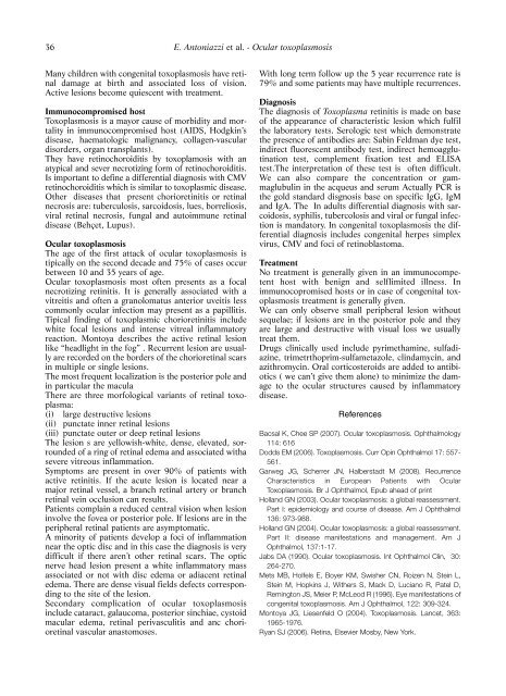
Many children with congenital toxoplasmosis have retinal<br />
damage at birth and associated loss of vision.<br />
Active lesions become quiescent with treatment.<br />
Immunocompromised host<br />
Toxoplasmosis is a mayor cause of morbi<strong>di</strong>ty and mortality<br />
in immunocompromised host (AIDS, Hodgkin’s<br />
<strong>di</strong>sease, haematologic malignancy, collagen-vascular<br />
<strong>di</strong>sorders, organ transplants).<br />
They have retinochoroi<strong>di</strong>tis by toxoplamosis with an<br />
atypical and sever necrotizing form of retinochoroi<strong>di</strong>tis.<br />
Is important to define a <strong>di</strong>fferential <strong>di</strong>agnosis with CMV<br />
retinochoroi<strong>di</strong>tis which is similar to toxoplasmic <strong>di</strong>sease.<br />
Other <strong>di</strong>seases that present chorioretinitis or retinal<br />
necrosis are: tuberculosis, sarcoidosis, lues, borreliosis,<br />
viral retinal necrosis, fungal and autoimmune retinal<br />
<strong>di</strong>sease (Behçet, Lupus).<br />
Ocular toxoplasmosis<br />
The age of the first attack of ocular toxoplasmosis is<br />
tipically on the second decade and 75% of cases occur<br />
between 10 and 35 years of age.<br />
Ocular toxoplasmosis most often presents as a focal<br />
necrotizing retinitis. It is generally associated with a<br />
vitreitis and often a granolomatus anterior uveitis less<br />
commonly ocular infection may present as a papillitis.<br />
Tipical fin<strong>di</strong>ng of toxoplasmic chorioretinitis include<br />
white focal lesions and intense vitreal inflammatory<br />
reaction. Montoya describes the active retinal lesion<br />
like “headlight in the fog” . Recurrent lesion are usually<br />
are recorded on the borders of the chorioretinal scars<br />
in multiple or single lesions.<br />
The most frequent localization is the posterior pole and<br />
in particular the macula<br />
There are three morfological variants of retinal toxoplasma:<br />
(i) large destructive lesions<br />
(ii) punctate inner retinal lesions<br />
(iii) punctate outer or deep retinal lesions<br />
The lesion s are yellowish-white, dense, elevated, sorrounded<br />
of a ring of retinal edema and associated witha<br />
severe vitreous inflammation.<br />
Symptoms are present in over 90% of patients with<br />
active retinitis. If the acute lesion is located near a<br />
major retinal vessel, a branch retinal artery or branch<br />
retinal vein occlusion can results.<br />
Patients complain a reduced central vision when lesion<br />
involve the fovea or posterior pole. If lesions are in the<br />
peripheral retinal patients are asymptomatic.<br />
A minority of patients develop a foci of inflammation<br />
near the optic <strong>di</strong>sc and in this case the <strong>di</strong>agnosis is very<br />
<strong>di</strong>fficult if there aren’t other retinal scars. The optic<br />
nerve head lesion present a white inflammatory mass<br />
associated or not with <strong>di</strong>sc edema or a<strong>di</strong>acent retinal<br />
edema. There are dense visual fields defects correspon<strong>di</strong>ng<br />
to the site of the lesion.<br />
Secondary complication of ocular toxoplasmosis<br />
include cataract, galaucoma, posterior sinchiae, cystoid<br />
macular edema, retinal perivasculitis and anc chorioretinal<br />
vascular anastomoses.<br />
E. Antoniazzi et al. - Ocular toxoplasmosis<br />
With long term follow up the 5 year recurrence rate is<br />
79% and some patients may have multiple recurrences.<br />
Diagnosis<br />
The <strong>di</strong>agnosis of Toxoplasma retinitis is made on base<br />
of the appearance of characteristic lesion which fulfil<br />
the laboratory tests. Serologic test which demonstrate<br />
the presence of antibo<strong>di</strong>es are: Sabin Feldman dye test,<br />
in<strong>di</strong>rect fluorescent antibody test, in<strong>di</strong>rect hemoagglutination<br />
test, complement fixation test and ELISA<br />
test.The interpretation of these test is often <strong>di</strong>fficult.<br />
We can also compare the concentration or gammaglubulin<br />
in the acqueus and serum Actually PCR is<br />
the gold standard <strong>di</strong>sgnosis base on specific IgG, IgM<br />
and IgA. The In adults <strong>di</strong>fferential <strong>di</strong>agnosis with sarcoidosis,<br />
syphilis, tubercolosis and viral or fungal infection<br />
is mandatory. In congenital toxoplasmosis the <strong>di</strong>fferential<br />
<strong>di</strong>agnosis includes congenital herpes simplex<br />
virus, CMV and foci of retinoblastoma.<br />
Treatment<br />
No treatment is generally given in an immunocompetent<br />
host with benign and selflimited illness. In<br />
immunocopromised hosts or in case of congenital toxoplasmosis<br />
treatment is generally given.<br />
We can only observe small peripheral lesion without<br />
sequelae; if lesions are in the posterior pole and they<br />
are large and destructive with visual loss we usually<br />
treat them.<br />
Drugs clinically used include pyrimethamine, sulfa<strong>di</strong>azine,<br />
trimetrthoprim-sulfametazole, clindamycin, and<br />
azithromycin. Oral corticosteroids are added to antibiotics<br />
( we can’t give them alone) to minimize the damage<br />
to the ocular structures caused by inflammatory<br />
<strong>di</strong>sease.<br />
References<br />
Bacsal K, Chee SP (2007). Ocular toxoplasmosis. Ophthalmology<br />
114: 616<br />
Dodds EM (2006). Toxoplasmosis. Curr Opin Ophthalmol 17: 557-<br />
561.<br />
Garweg JG, Scherrer JN, Halberstadt M (2008). Recurrence<br />
Characteristics in European Patients with Ocular<br />
Toxoplasmosis. Br J Ophthalmol, Epub ahead of print<br />
Holland GN (2003). Ocular toxoplasmosis: a global reassessment.<br />
Part I: epidemiology and course of <strong>di</strong>sease. Am J Ophthalmol<br />
136: 973-988.<br />
Holland GN (2004). Ocular toxoplasmosis: a global reassessment.<br />
Part II: <strong>di</strong>sease manifestations and management. Am J<br />
Ophthalmol, 137:1-17.<br />
Jabs DA (1990). Ocular toxoplasmosis. Int Ophthalmol Clin, 30:<br />
264-270.<br />
Mets MB, Holfels E, Boyer KM, Swisher CN, Roizen N, Stein L,<br />
Stein M, Hopkins J, Withers S, Mack D, Luciano R, Patel D,<br />
Remington JS, Meier P, McLeod R (1996). Eye manifestations of<br />
congenital toxoplasmosis. Am J Ophthalmol, 122: 309-324.<br />
Montoya JG, Liesenfeld O (2004). Toxoplasmosis. Lancet, 363:<br />
1965-1976.<br />
Ryan SJ (2006). Retina, Elsevier Mosby, New York.