impaginato piccolo - Società Italiana di Parassitologia (SoIPa)
impaginato piccolo - Società Italiana di Parassitologia (SoIPa)
impaginato piccolo - Società Italiana di Parassitologia (SoIPa)

Create successful ePaper yourself
Turn your PDF publications into a flip-book with our unique Google optimized e-Paper software.
<strong>Parassitologia</strong> 50: 93-95, 2008<br />
Uncommon cases of pityriasis versicolor<br />
C. Romano<br />
Dermatologic Section of the Department of Clinical Me<strong>di</strong>cine and Immunological Science. University of Siena, Italy<br />
Abstract. Malassezia may play a role in several dermatoses. It is responsible for foliculitis and mainly for<br />
pityriasis versicolor. Pityriasis versicolor is the most known dermatitis because of its clinical aspects and frequently<br />
for its poor response to the therapy, mainly in chronic forms. The clinical aspects of uncommon and<br />
rare forms of pityriasis versicolor have been reported. The data related to the patients observed in the last<br />
thirty years in Siena are reported. In ad<strong>di</strong>tion, a study was carried out in Pisa by Professor F. Mancianti to<br />
identify species of Malassezia isolated in 37 patients.<br />
Key words: Pityriasis versicolor, mycoses, Malassezia<br />
Malassezia is responsible for pityriasis versicolor (PV)<br />
and folliculitis, and it is hypothesized that play a role in<br />
the pathogenesis of seborrhoeic dermatitis (SD), and<br />
atopic dermatitis (AD). Nevertheless the most frequent<br />
dermatological <strong>di</strong>sease caused by this mycete is certainly<br />
the pityriasis versicolor, which commonly manifests<br />
with hyperpigmented or achromic macules, accor<strong>di</strong>ng<br />
to the season. Lesions are most frequent on the seborrhoeic<br />
areas, such as the upper trunk, neck and upperarms.<br />
Besides the typical manifestations, there are<br />
those ones which are uncommon due to their clinical<br />
aspects (pityriasis versicolor atrophicans and pityriasis<br />
versicolor rubra) or due to their sites (pytiriasis versicolor<br />
areolar e periareolar, penile, in the groins, perianal,<br />
palmar and plantar areas). There are even forms<br />
thet affect almost all body surface.<br />
Crowson and coll. reported twelve clinical cases in<br />
which lesions characterized as patches, macules, and<br />
atrophic plaques prompted clinical <strong>di</strong>fferential <strong>di</strong>agnosis<br />
with atrophy due to intralesional steroid therapy,<br />
collagen <strong>di</strong>seases or parapsoriasis. However, histologic<br />
examination showed hyphae and spores in the corneum<br />
layer, variable epidermal and dermal atrophies, reteridge<br />
effacement, subepidermal fibroplasia, pigment<br />
incontinence and elastolysis.This form was defined<br />
atrophying pitiriasis versicolor (1, 2).<br />
Pitiriasis versicolor rubra clinically presented as soybean-sized,<br />
red colored macule. At first it was primarily<br />
reported by Horiuchi, and recently six cases attributed<br />
to Malassezia sympo<strong>di</strong>alis have been described.<br />
Rudolph and coll. described two patients who had<br />
some lightly pigmented, scaling, mildly pruritic eruptions<br />
in the groins, axillae, and perianal regions. The<br />
<strong>di</strong>fferential <strong>di</strong>agnosis included erythrasma, SD and dermatophyte<br />
infections. The authors have stressed a new<br />
nomenclature, “inverse tinea versicolor”, due to the<br />
highly atypical site.<br />
Correspondence: Clara Romano<br />
Dept. of Clinical Me<strong>di</strong>cine and Immunological Science,<br />
Section of Dermatology, University of Siena,<br />
Policlinico le Scotte, Viale Bracci 53100 Siena, Italy.<br />
e-mail: romanoclara@unisi.it<br />
Anthony reported circular areas roughly hypopigmented,<br />
unilateral or bilateral with thin scales localized in<br />
the areolae or in the periareolae.<br />
Schosser and coll. reported the case of man who had a<br />
brownish, scaly lesion on the nipple microscopically<br />
<strong>di</strong>agnosed as areolar and periareolar PV.<br />
Kamalam showed similar lesions in the penis, in the<br />
groins, in the perianal, palmar and plantar areas.<br />
Blumenthal described multiple hypopigmented and<br />
scaly macular lesions over the penis of a patient who<br />
has also been treated during the last five years with triamcinolone<br />
acetonide and prednisolone acetate for<br />
neurodermatitis, which was not the correct <strong>di</strong>agnosis.<br />
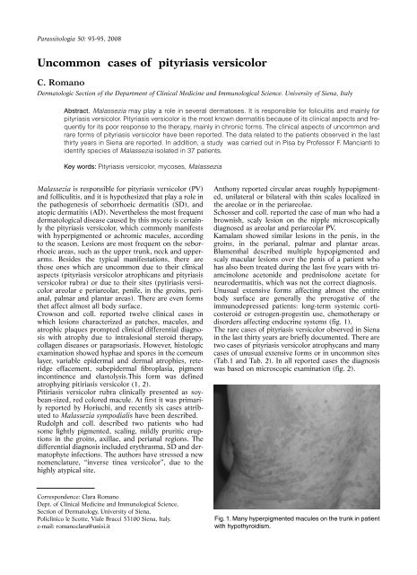
Unusual extensive forms affecting almost the entire<br />
body surface are generally the prerogative of the<br />
immunodepressed patients: long-term systemic corticosteroid<br />
or estrogen-progestin use, chemotherapy or<br />
<strong>di</strong>sorders affecting endocrine systems (fig. 1).<br />
The rare cases of pityriasis versicolor observed in Siena<br />
in the last thirty years are briefly documented. There are<br />
two cases of pityriasis versicolor atrophycans and many<br />
cases of unusual extensive forms or in uncommon sites<br />
(Tab.1 and Tab. 2). In all reported cases the <strong>di</strong>agnosis<br />
was based on microscopic examination (fig. 2).<br />
Fig. 1. Many hyperpigmented macules on the trunk in patient<br />
with hypothyroi<strong>di</strong>sm.