View Annual Report - Jules Stein Eye Institute
View Annual Report - Jules Stein Eye Institute
View Annual Report - Jules Stein Eye Institute

You also want an ePaper? Increase the reach of your titles
YUMPU automatically turns print PDFs into web optimized ePapers that Google loves.
Clinical Laboratories<br />
The Ophthalmology Clinical Laboratories provide<br />
precise measurements, photographs, and quantitative<br />
studies of the eye and the visual system. Quantitative<br />
information of this type enhances patient care by<br />
increasing the accuracy of diagnosis and enlarging<br />
the parameters employed to assess the clinical course<br />
and effectiveness of treatment. Additionally, the clinical<br />
laboratories expand the scope of treatment alternatives,<br />
promote clinical research, and generally augment the<br />
effectiveness of ophthalmic disease management.<br />
The laboratories are available to all ophthalmologists<br />
in the community.<br />
Corneal Diagnostic Laboratory<br />
The Corneal Diagnostic Laboratory, under the direction<br />
of Dr. Anthony J. Aldave, offers a comprehensive array<br />
of corneal imaging modalities. Services include imaging<br />
of the anterior and posterior corneal surfaces with the<br />
Marco OPD-Scan III and Bausch and Lomb Orbscan<br />
topographers and the Ziemer GALILEI Dual Scheimpflug<br />
Analyzer, and imaging of the corneal endothelium for<br />
assessment of corneal endothelial cell morphology<br />
and density using the KONAN CellChek XL specular<br />
microscope. Full-thickness confocal microscopic<br />
imaging of the cornea, a very useful tool in the diagnosis<br />
of suspected fungal, acanthamoebic and other<br />
parasitic infections of the cornea, is performed with<br />
the Heidelberg HRT3 confocal microscope. This<br />
instrument can also perform optical pachymetry to<br />
noninvasively measure LASIK residual bed thicknesses<br />
and flap thicknesses as well as evaluate the LASIK<br />
interface for possible infections, diffuse lamellar<br />
keratitis, and ingrowth.<br />
84 Programs | Clinical Laboratories<br />
Glaucoma Photography Laboratory<br />
The Glaucoma Photography Laboratory, under the<br />
direction of Dr. Joseph Caprioli, provides specialized<br />
photographs for new and follow-up glaucoma patients<br />
to assist the ophthalmologist in the management of<br />
patients with this disease. The GDX Nerve Fiber<br />
Analyzer utilizes polarized light in place of dilation to<br />
measure the thickness of the nerve fiber layer. This<br />
test is particularly useful in diagnosing new glaucoma.<br />
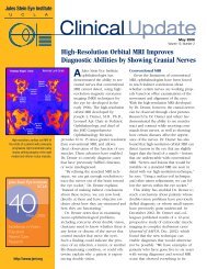
Heidelberg retinal tomography, using confocal laser<br />
light, measures additional parameters of the optic nerve,<br />
and provides more information on the nerve fiber layer.<br />
Optical coherence tomography utilizes reflected light<br />
to measure the nerve fiber layer as well as to measure<br />
macular holes as a staging procedure for surgical repair.<br />
An ophthalmic fundus camera photographs the optic<br />
nerve in stereo. The Laboratory is conducting clinical<br />
studies to evaluate the effectiveness of each photographic<br />
modality in terms of predictive accuracy and<br />
early detection of glaucoma.<br />
Ocular Motility<br />
Clinical and Basic Science Laboratory<br />
The Ocular Motility Clinical and Basic Science Laboratory,<br />
under the direction of Dr. Joseph L. Demer,<br />
records and quantitatively analyzes eye movement<br />
abnormalities resulting from ocular and neurological<br />
disorders, such as ocular myasthenia gravis. Several<br />
types of tests are performed. The Hess test utilizes<br />
specialized eye charts and lenses to assist in the diag-<br />
nosis of a number of problems, including double vision.<br />
Magnetic scleral search coil techniques are utilized in<br />
clinical research studies to detect fine movements not<br />
evident through normal visual examination. Another test<br />
involves the visual recording of eye movement using a<br />
video camera. The Laboratory also engages in basic<br />
science research to further understanding of eye<br />
movement as well as diseases of the eye, brain, and<br />
muscles, and related tissues of the inner ear.<br />
Under the direction of Dr. Joseph L. Demer<br />
(standing), the Ocular Motility Clinical and<br />
Basic Science Laboratory engages in basic<br />
science research to further understanding<br />
of eye movement and diseases of the eye,<br />
brain, muscles, and related tissues of the<br />
inner ear.