View Annual Report - Jules Stein Eye Institute
View Annual Report - Jules Stein Eye Institute
View Annual Report - Jules Stein Eye Institute

Create successful ePaper yourself
Turn your PDF publications into a flip-book with our unique Google optimized e-Paper software.
Ophthalmic Photography<br />
Clinical Laboratory<br />
The Ophthalmic Photography Clinical Laboratory, under<br />
the direction of Dr. Tara A. McCannel, provides a wide<br />
array of photographic techniques important in patient<br />
care, research, and teaching. The primary purpose of<br />
ophthalmic photography in patient care is to record the<br />
present state of the eye, and in cases of abnormality, to<br />
establish a baseline and monitor the patient’s condition<br />
over time. Patient care services include photographic<br />
documentation of anterior segment diseases involving<br />
corneal problems like growths, infection, and trauma;<br />
photographs of ocular motility to record abnormalities<br />
in eye movement; fundus photography, which captures<br />
pictures of the retina; and diagnostic testing using<br />
fluorescein and indocyanine green angiography, which<br />
records the dynamics of blood flow in the eye. The<br />
Laboratory also supports the research and teaching<br />
activities of the <strong>Jules</strong> <strong>Stein</strong> <strong>Eye</strong> <strong>Institute</strong> by preparing<br />
and duplicating graphic materials for presentation<br />
and publication.<br />
Ophthalmic Ultrasonography<br />
Clinical Laboratory<br />
The Ophthalmic Ultrasonography Clinical Laboratory,<br />
directed by Dr. Steven D. Schwartz, performs clinical<br />
examinations that are useful in diagnosing both ocular<br />
and orbital eye diseases. Diagnostic examinations<br />
include standardized A-scan, B-scan, and biomicroscopy.<br />
Standardized A-scan is useful in tissue differentiation<br />
and is commonly employed to diagnose ocular and<br />
orbital tumors, including choroidal melanoma. B-scan<br />
provides location and contour information and is par-<br />
ticularly useful in differentiating vitreous membranes<br />
from retinal detachment. Ultrasound biomicroscopy<br />
provides exquisitely detailed, high-resolution views of<br />
the anterior segment of the eye and is a critical tool for<br />
the evaluation of ocular pathology, especially in opaque<br />
corneas. Biometry and intraocular lens calculations are<br />
also performed in the Laboratory, under the direction of<br />
Dr. Ralph Levinson. Biometry measures the axial eye<br />
length, anterior chamber depth, and lens thickness;<br />
intraocular lens calculations are performed to determine<br />
the power of the lens implant for cataract patients.<br />
Perimetry Laboratory<br />
The Perimetry Laboratory, under the direction of<br />
Dr. Joseph Caprioli, performs visual field examinations<br />
that determine the sensitivity of central and peripheral<br />
vision. Examinations are conducted with advanced<br />
Humphrey automated perimetry equipment. Testing<br />
detects visual field deficits associated with certain kinds<br />
of eye diseases such as glaucoma, retinal disorders,<br />
and neuro-ophthalmic conditions. Utilizing pinpoints<br />
of light around a perimetry bowl, the test evaluates<br />
different areas of the field of vision. Test results are<br />
computerized and compared to a range of normal<br />
values by age group. Patterns of diminished fields of<br />
vision are related to specific eye diseases. Perimetry<br />
testing is employed for diagnostic purposes and to<br />
monitor visual field sensitivity over time, especially for<br />
glaucoma patients. Both standard and shortwave auto-<br />
mated techniques are available, in addition to frequencydoubling<br />
perimetry and motion-detection perimetry.<br />
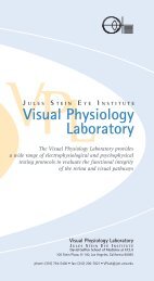
Visual Physiology Clinical Laboratory<br />
The Visual Physiology Clinical Laboratory, under<br />
the direction of Drs. Michael B. Gorin and Steven<br />
Nusinowitz, quantitatively evaluates the function of<br />
the retina and visual pathways. Patients are referred<br />
for functional testing to confirm a specific diagnosis<br />
or, in cases where the etiology is unknown, to rule out<br />
alternative diagnostic possibilities. Electrophysiological<br />
tests, including both the full-field and multifocal electroretinograms<br />
(ERG and mfERG), the electro-oculogram<br />
(EOG), and visually evoked cortical potentials (VECP),<br />
record electrical signals from different layers of the<br />
visual system to identify the site responsible for visual<br />
symptoms. Psychophysical tests require the participation<br />
of the patient in specific tasks to evaluate visual<br />
functions like color blindness, contrast sensitivity, and<br />
visual acuity. In many cases, both electrophysical and<br />
psychophysical tests are performed together to obtain<br />
the optimum amount of information for diagnosis.<br />
Programs | Clinical Laboratories 85