THIS WEEK IN
THIS WEEK IN
THIS WEEK IN

Create successful ePaper yourself
Turn your PDF publications into a flip-book with our unique Google optimized e-Paper software.
much less efficiently in the ipk1D strain (Fig. 6C).<br />
A37 in the wild-type strain is read as a thymidine,<br />
presumably due to N 1 -methylinosine at position<br />
37. m 1 I, like m 1 A, may pair with reduced specificity<br />
in the reverse transcription reaction, explaining<br />
the presence of a T in the PCR product (36).<br />
In the crystal structure of hADAR2, IP 6<br />
binds<br />
and fills an extremely basic hole, with the center<br />
of the inositol ring more than 10 ) from the<br />
protein surface. Thus, it seems possible that<br />
ADAR2 and, by analogy, ADAT1, are unstable<br />
without IP 6<br />
. In this regard we wondered about the<br />
nature of ADAT1 expressed in the ipk1D mutant.<br />
Was this protein trapped in an irreversible inactive<br />
state or forming a folding intermediate that<br />
couldbindIP 6<br />
to achieve its active conformation?<br />
To explore this question, we tested whether the<br />
addition of IP 6<br />
to extracts prepared from the<br />
ipk1D mutant could recover ADAT1 activity.<br />
When added to extracts prepared from the ipk1D<br />
strain, IP 6<br />
recovered activity to È50% of wildtype<br />
(Fig. 7, A and C), which suggests that the<br />
protein does not require IP 6<br />
during its synthesis<br />
and the initial stages of folding. As expected, the<br />
addition of IP 6<br />
to wild-type extract had no effect,<br />
because these cells are capable of synthesizing<br />
IP 6<br />
(Fig. 7, B and C). To test for the specific<br />
requirement for IP 6<br />
by ADAT1, we performed a<br />
similar experiment, except we substituted inositol<br />
hexakissulfate (IS 6<br />
)forIP 6<br />
. Despite its similar<br />
charge and structure, IS 6<br />
does not recover<br />
ADAT1 activity when added to ipk1D extracts<br />
(Fig. 7A). This suggests that the enzyme specifically<br />
requires IP 6<br />
for function and can discriminate<br />
between the minor differences in phosphate/<br />
sulfate chemistry (e.g., the protonation state).<br />
So far, we have been unable to rescue the<br />
activity of hADAR2 expressed in the ipk1D<br />
by adding IP 6<br />
. Possibly, native S. cerevisiae<br />
ADAT1, but not the heterologous hADAR2, is<br />
associated with a host chaperone in the extract<br />
that promotes refolding in the presence of IP 6<br />
.<br />
Alternatively, this result may hint at interesting<br />
differences between the two enzymes in IP 6<br />
accessibility.<br />
Such a difference might explain why<br />
assays of ADAT1 in ipk1D extracts show a small<br />
(È5%) amount of A37 deamination at the highest<br />
concentrations of extract (Fig. 6A), whereas<br />
ADAR2 expressed in this strain shows no residual<br />
activity. If the IP 6<br />
binding site in ADAT1<br />
were more accessible than that of ADAR2, it<br />
might bind a noncognate inositol polyphosphate,<br />
such as IP 5<br />
, to allow a low level of activity.<br />
Discussion. Burial of IP 6<br />
may reflect a novel<br />
way of using an available cellular component<br />
R ESEARCH A RTICLE<br />
to define and stabilize a protein fold. This would<br />
be analogous to the use of Bstructural[ metal<br />
ions in stabilizing the fold of metalloproteins.<br />
To our knowledge, this represents the first example<br />
of a protein using IP 6<br />
for this purpose. Other<br />
protein structures with bound IP 6<br />
have been<br />
reported, such as deoxyhemoglobin (37) andthe<br />
clathrin adaptor complex AP2 (38); however,<br />
unlike ADAR2-D, in these cases the IP 6<br />
molecule<br />
is not extensively buried and does not appear<br />
to dramatically stabilize the overall structure.<br />
In addition to the structural requirement, IP 6<br />
may play a subtle role in modulating catalytic<br />
efficiency by indirectly ordering the side chain<br />
of K483. Two of the IP 6<br />
phosphate groups approach<br />
within 10 ) of the catalytic zinc ion and<br />
are indirectly coordinated to zinc by a chain of<br />
hydrogen-bonded residues that includes K519,<br />
D392, and K483 (Fig. 3A). These residues are<br />
conserved among active ADARs, and K483 may<br />
contribute to tuning the pK a<br />
of the nucleophilic<br />
water molecule through its interaction with C516<br />
(Fig. 3A).<br />
Sequence alignments indicate that ADAT1<br />
enzymes are the evolutionary link between the<br />
other family members, ADAT2/3 (including<br />
TadA) and ADARs (18). ADAT1 apparently<br />
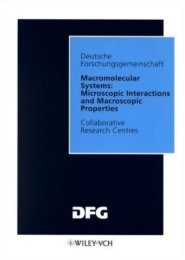
Fig. 4. (A) The 27-mer R/G site RNA substrate<br />
used to assay hADAR2 editing activity. (B) Editing<br />
of the R/G site RNA by hADAR2 expressed in<br />
wild-type or ipk1D yeast. The R/G site adenosine<br />
was labeled with 32 P and incubated with increasing<br />
concentrations of expressed hADAR2 in<br />
extracts. Reacted RNA was treated with nuclease<br />
P1, the resulting 5 nucleotide monophosphates<br />
separated by thin-layer chromatography (TLC),<br />
and the plate exposed to a PhosphorImager<br />
screen. The amount of hADAR2 in each extract<br />
was determined by Western blotting, and extract<br />
was added to give the final ADAR2 concentrations<br />
as indicated. (C) Western blot showing the<br />
amount of hADAR2 in each reaction. Single-letter<br />
abbreviations for amino acid residues are defined<br />
in (42).<br />
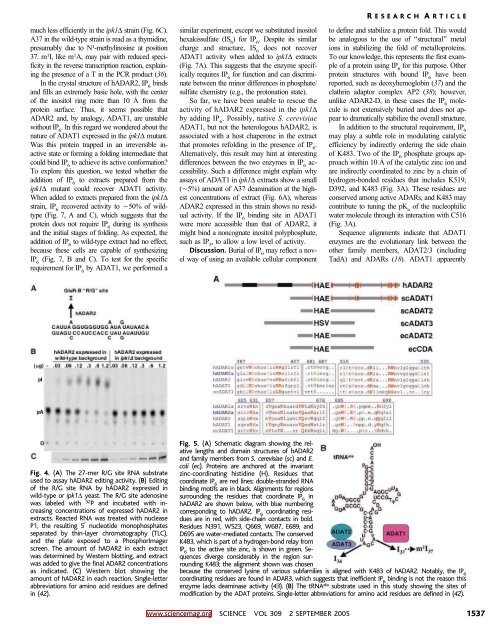
Fig. 5. (A) Schematic diagram showing the relative<br />
lengths and domain structures of hADAR2<br />
and family members from S. cerevisiae (sc) and E.<br />
coli (ec). Proteins are anchored at the invariant<br />
zinc-coordinating histidine (H). Residues that<br />
coordinate IP 6<br />
are red lines; double-stranded RNA<br />
binding motifs are in black. Alignments for regions<br />
surrounding the residues that coordinate IP 6<br />
in<br />
hADAR2 are shown below, with blue numbering<br />
corresponding to hADAR2. IP 6<br />
coordinating residues<br />
are in red, with side-chain contacts in bold.<br />
Residues N391, W523, Q669, W687, E689, and<br />
D695 are water-mediated contacts. The conserved<br />
K483, which is part of a hydrogen-bond relay from<br />
IP 6<br />
to the active site zinc, is shown in green. Sequences<br />
diverge considerably in the region surrounding<br />
K483; the alignment shown was chosen<br />
because the conserved lysine of various subfamilies is aligned with K483 of hADAR2. Notably, the IP 6<br />
coordinating residues are found in ADAR3, which suggests that inefficient IP 6<br />
binding is not the reason this<br />
enzyme lacks deaminase activity (43). (B) ThetRNA ala substrate used in this study showing the sites of<br />
modification by the ADAT proteins. Single-letter abbreviations for amino acid residues are defined in (42).<br />
www.sciencemag.org SCIENCE VOL 309 2 SEPTEMBER 2005 1537