THIS WEEK IN
THIS WEEK IN
THIS WEEK IN

You also want an ePaper? Increase the reach of your titles
YUMPU automatically turns print PDFs into web optimized ePapers that Google loves.
RNA<br />
S PECIAL S ECTION<br />
rapidly, with new miRNA genes arising by<br />
duplication and mutation of the 21-nt miRNA<br />
sequence.<br />
Small on Specificity<br />
From the standpoint of binding specificity, small<br />
RNAs are truly diminutive. A mere six or seven<br />
of the 21 nucleotides within a miRNA or siRNA<br />
provide the bulk of binding specificity for the<br />
small RNA–protein complexes they guide. As<br />
first proposed by Lai (29), and subsequently<br />
confirmed computationally (3, 30) and experimentally<br />
for miRNAs (31–34) and siRNAs<br />
(35, 36), the 5 end of a miRNA or an siRNA<br />
contributes disproportionately to target RNA<br />
binding. Kinetic and structural studies suggest<br />
that the first nucleotide of a small RNA guide is<br />
unpaired during small RNA function (36–38).<br />
The small region of the small RNA that<br />
mediates target binding has been called the<br />
‘‘seed sequence,’’ a term intended to suggest<br />
that the region nucleates binding between the<br />
small RNA guide and its target, and that the<br />
more 3 regions of the small RNA subsequently<br />
zipper-up—if they can—with the 5 regions<br />
of the binding site on the target RNA (39).<br />
In truth, current experimental evidence<br />
cannot discern the order in which distinct<br />
regions of the small RNA interact with its<br />
binding site on the target RNA. Both computational<br />
and experimental approaches detect<br />
only the binding contributions of specific small<br />
RNA regions at equilibrium. But the finding<br />
that stable binding between the small RNA<br />
and its target derives from such a small region<br />
of an already puny RNA oligonucleotide<br />
implies that the manner in which the small<br />
RNA interacts with its target is very different<br />
from antisense oligonucleotide–target RNA<br />
pairing. This radical and unexpected mode of<br />
nucleic acid interaction is almost certainly a<br />
consequence of the way the small RNA—both<br />
alone and paired to its RNA target—is bound<br />
by a member of the Argonaute family of proteins.<br />
These multidomain proteins are specialized<br />
for binding the small RNAs that<br />
mediate RNA silencing; understanding the<br />
relationship of Argonaute protein structure to<br />
their functions in controlling gene expression<br />
is now the key to understanding the deeper<br />
physical meaning of the small RNA ‘‘seed<br />
sequence.’’<br />
The small RNAs that act in RNA silencing<br />
pathways are like fancy restriction enzymes<br />
whose recognition sites occur at random once<br />
every È4000 to È65,000 nt of sequence. But<br />
unlike restriction enzymes, which cut DNA<br />
wherever they bind, small RNAs can act in two<br />
distinct ways, each of which dramatically<br />
extends their functional specificity (Fig. 3) (2).<br />
When a small RNA pairs extensively with its<br />
RNA target, it directs cleavage of a single<br />
phosphodiester bond in the target RNA, across<br />
from nucleotides 10 and 11 of the small RNA<br />
guide (40). Thus, small RNA–directed cleavage<br />
is much more specific than small RNA binding<br />
itself, as it occurs only when most of the 21 nt<br />
of the siRNA or miRNA can base pair to<br />
form at least one turn of an A-form helix<br />
with the RNA target (36, 41, 42). Even when<br />
the small RNA is fully complementary to its<br />
target RNA, cleavage only occurs when the<br />
RNA is bound to the right Argonaute protein<br />
(43, 44). In humans, only one of the four<br />
Argonaute proteins examined in detail retains<br />
all the amino acids required to catalyze target<br />
RNA cleavage (45). Argonaute proteins contain<br />
two RNA-binding domains: the Piwi<br />
domain, which binds the small RNA guide at<br />
its 5 end, and the PAZ domain, which binds<br />
the single-stranded 3 end of small RNA. The<br />
endonuclease that cleaves target RNAs resides<br />
in the Piwi domain, and this domain is a<br />
structural homolog of the DNA-guided RNA<br />
endonuclease RNase H (46). Target RNA<br />
cleavage is commonly viewed as the siRNA<br />
or RNAi mode, but is actually the dominant<br />
mechanism by which plant miRNAs regulate<br />
their targets (47, 48) and is found for at least<br />
a small number of animal (49, 50) and viral<br />
miRNAs (51).<br />
In Drosophila or human cell lysates, small<br />
RNA–programmed Argonaute2 (Ago2) acts as<br />
a multiple-turnover enzyme, with each small<br />
RNA directing the cleavage of hundreds of<br />
target molecules (36, 52). Small RNA–directed<br />
mRNA cleavage cuts an mRNA into two<br />
pieces, and efficient release of these fragments<br />
requires adenosine triphosphate (ATP) (36).<br />
Proteins besides Ago2 may be required for<br />
release of the products of small RNA–directed<br />
target cleavage. In fact, Ago2 alone can direct<br />
a single round of target cleavage but cannot<br />
efficiently catalyze additional cycles, likely<br />
because the cleavage products remain bound<br />
to the small RNA within the enzyme (45).<br />
After the cleaved pieces of the target are<br />
released, the 3 fragment is destroyed in the<br />
cytoplasm by the exonuclease Xrn1 while the<br />
5 fragment is degraded by the exosome, a<br />
collection of exonucleases dedicated to 3-to-5<br />
RNA degradation (53). In plants and animals,<br />
when miRNAs direct mRNA cleavage, a short<br />
polyuridine [poly(U)] tail is subsequently<br />
addedtothe3 end of the 5 cleavage fragment<br />
(54). Addition of poly(U) correlates with decapping<br />
and 5-to-3 destruction of the target<br />
RNA cleavage fragment, at least in plants,<br />
suggesting an alternative route to the exosome<br />
for degradation of the 5 cleavage product.<br />
When siRNAs or miRNAs pair only partially<br />
with their targets, they cannot direct<br />
mRNA cleavage. Instead, they block translation<br />
of the mRNA into protein (55, 56). However,<br />
binding of a single miRNA alone is<br />
usually insufficient to measurably block<br />
translation; instead, several miRNAs bind to<br />
the same target—opening the door to combinatorial<br />
control of gene expression by sets of<br />
coordinately expressed miRNAs (39). Initially,<br />
miRNAs were proposed to repress translation<br />
at a step after ribosomes have bound<br />
the mRNA, i.e., after translational initiation<br />
(55). One idea was that they direct degradation<br />
of the nascent polypeptide as it emerges<br />
from the ribosome. Alternatively, they might<br />
‘‘freeze’’ ribosomes in place on the mRNA,<br />
stalling elongation of the growing protein<br />
chain. Recent findings, however, call these<br />
ideas into question. For example, translational<br />
repression by miRNAs was thought to affect<br />
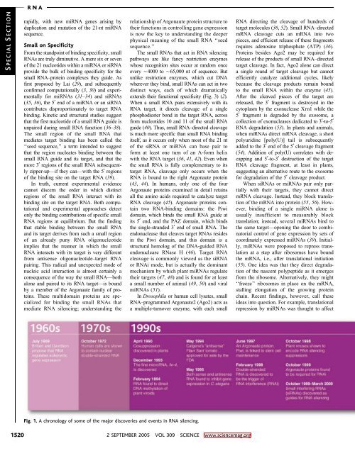
Fig. 1. A chronology of some of the major discoveries and events in RNA silencing.<br />
1520<br />
2 SEPTEMBER 2005 VOL 309 SCIENCE www.sciencemag.org