THIS WEEK IN
THIS WEEK IN
THIS WEEK IN

Create successful ePaper yourself
Turn your PDF publications into a flip-book with our unique Google optimized e-Paper software.
R EPORTS<br />
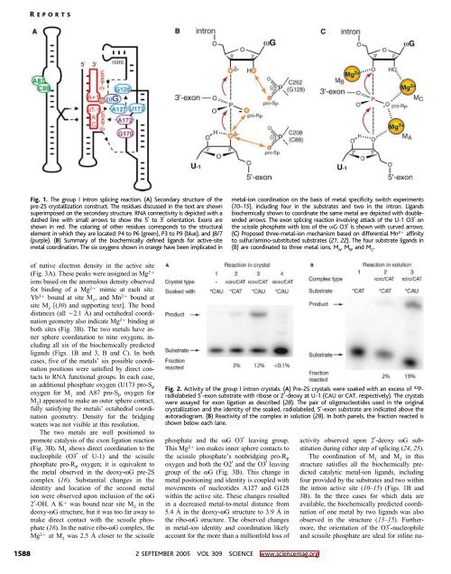
Fig. 1. The group I intron splicing reaction. (A) Secondary structure of the<br />
pre-2S crystallization construct. The residues discussed in the text are shown<br />
superimposed on the secondary structure. RNA connectivity is depicted with a<br />
dashed line with small arrows to show the 5 to 3 orientation. Exons are<br />
shown in red. The coloring of other residues corresponds to the structural<br />
element in which they are located: P4 to P6 (green), P3 to P9 (blue), and J8/7<br />
(purple). (B) Summary of the biochemically defined ligands for active-site<br />
metal coordination. The six oxygens shown in orange have been implicated in<br />
metal-ion coordination on the basis of metal specificity switch experiments<br />
(10–15), including four in the substrates and two in the intron. Ligands<br />
biochemically shown to coordinate the same metal are depicted with doubleended<br />
arrows. The exon splicing reaction involving attack of the U-1 O3 on<br />
the scissile phosphate with loss of the wG O3 is shown with curved arrows.<br />
(C) Proposed three-metal-ion mechanism based on differential Mn 2þ affinity<br />
to sulfur/amino-substituted substrates (21, 22). The four substrate ligands in<br />
(B) are coordinated to three metal ions, M A<br />
,M B<br />
,andM C<br />
.<br />
of native electron density in the active site<br />
(Fig. 3A). These peaks were assigned as Mg 2þ<br />
ions based on the anomalous density observed<br />
for binding of a Mg 2þ mimic at each site.<br />
Yb 3þ bound at site M 1<br />
,andMn 2þ bound at<br />
site M 2<br />
E(30) and supporting text^. The bond<br />
distances (all È2.1 )) and octahedral coordination<br />
geometry also indicate Mg 2þ binding at<br />
both sites (Fig. 3B). The two metals have inner<br />
sphere coordination to nine oxygens, including<br />
all six of the biochemically predicted<br />
ligands (Figs. 1B and 3, B and C). In both<br />
cases, five of the metals_ six possible coordination<br />
positions were satisfied by direct contacts<br />
to RNA functional groups. In each case,<br />
an additional phosphate oxygen (U173 pro-S P<br />
oxygen for M 1<br />
and A87 pro-S P<br />
oxygen for<br />
M 2<br />
) appeared to make an outer sphere contact,<br />
fully satisfying the metals_ octahedral coordination<br />
geometry. Density for the bridging<br />
waters was not visible at this resolution.<br />
The two metals are well positioned to<br />
promote catalysis of the exon ligation reaction<br />
(Fig.3B).M 1<br />
shows direct coordination to the<br />
nucleophile (O3 of U-1) and the scissile<br />
phosphate pro-R P<br />
oxygen; it is equivalent to<br />
the metal observed in the deoxy-wG pre-2S<br />
complex (16). Substantial changes in the<br />
identity and location of the second metal<br />
ion were observed upon inclusion of the wG<br />
2-OH. A K þ was bound near site M 2<br />
in the<br />
deoxy-wG structure, but it was too far away to<br />
make direct contact with the scissile phosphate<br />
(16). In the native ribo-wG complex,the<br />
Mg 2þ at M 2<br />
was 2.5 ) closer to the scissile<br />
Fig. 2. Activity of the group I intron crystals. (A) Pre-2Scrystalsweresoakedwithanexcessof 32 P-<br />
radiolabeled 5-exon substrate with ribose or 2-deoxy at U-1 (CAU or CAT, respectively). The crystals<br />
were assayed for exon ligation as described (28). The pair of oligonucleotides used in the original<br />
crystallization and the identity of the soaked, radiolabeled, 5-exon substrate are indicated above the<br />
autoradiogram. (B) Reactivity of the complex in solution (28). In both panels, the fraction reacted is<br />
shown below each lane.<br />
phosphate and the wG O3 leaving group.<br />
This Mg 2þ ion makes inner sphere contacts to<br />
the scissile phosphate_s nonbridging pro-R P<br />
oxygen and both the O2 and the O3 leaving<br />
group of the wG (Fig. 3B). This change in<br />
metal positioning and identity is coupled with<br />
movements of nucleotides A127 and G128<br />
within the active site. These changes resulted<br />
in a decreased metal-to-metal distance from<br />
5.4 ) in the deoxy-wG structure to 3.9 ) in<br />
the ribo-wG structure. The observed changes<br />
in metal-ion identity and coordination likely<br />
account for the more than a millionfold loss of<br />
activity observed upon 2-deoxy wG substitution<br />
during either step of splicing (24, 25).<br />
The coordination of M 1<br />
and M 2<br />
in this<br />
structure satisfies all the biochemically predicted<br />
catalytic metal-ion ligands, including<br />
four provided by the substrates and two within<br />
the intron active site (10–15) (Figs. 1B and<br />
3B). In the three cases for which data are<br />
available, the biochemically predicted coordination<br />
of one metal by two ligands was also<br />
observed in the structure (13–15). Furthermore,<br />
the orientation of the O3-nucleophile<br />
and scissile phosphate are ideal for inline nu-<br />
1588<br />
2 SEPTEMBER 2005 VOL 309 SCIENCE www.sciencemag.org