THIS WEEK IN
THIS WEEK IN
THIS WEEK IN

Create successful ePaper yourself
Turn your PDF publications into a flip-book with our unique Google optimized e-Paper software.
RNA<br />
plex, whereas the poly(A) tail carries a mixture of<br />
nuclear and cytoplasmic poly(A) binding proteins<br />
PABPN1 and PABPCs (Table 1). In this newly<br />
exported mRNP, CBC20/80 can functionally<br />
interact with translation initiation factor 4G<br />
(eIF4G), which serves to recruit the small<br />
ribosomal subunit and initiate 5Y3 scanning<br />
along the 5 UTR for an AUG start codon (27).<br />
Once the start codon is identified, the large<br />
ribosomal subunit is engaged to form an 80S<br />
complex competent for protein synthesis.<br />
Another major change in mRNP composition<br />
necessarily occurs upon the first passage of<br />
the 80S ribosome along the mRNA—the socalled<br />
‘‘pioneering round’’ of translation (28).<br />
Threading of the mRNA through the narrow<br />
space between the two ribosomal subunits<br />
strips away any remaining nuclear-acquired<br />
mRNP proteins, such as EJCs, residing inside<br />
the ORF. At some point, CBC20/80 and<br />
PABPN1 are also replaced by eIF4E (the<br />
major cytoplasmic cap-binding protein) and<br />
PABPCs, respectively. Whether these exchanges<br />
require any special mechanisms, such<br />
as the phosphorylation that promotes dissociation<br />
of Npl3p from newly exported mRNPs, or<br />
whether they occur simply as a consequence<br />
of mass action, is unknown. Regarding the<br />
second possibility, the low cytoplasmic concentrations<br />
of CBC20/80 and PABPN1<br />
coupled with the high concentrations of eIF4E<br />
and PABPCs could naturally lead to the latter<br />
set replacing the former, given reasonable dissociation<br />
rates. In any event, once the transition<br />
is complete, a network of simultaneous<br />
interactions between the 5 cap,eIF4E,eIF4G,<br />
PABPCs, and the poly(A) tail results in functional<br />
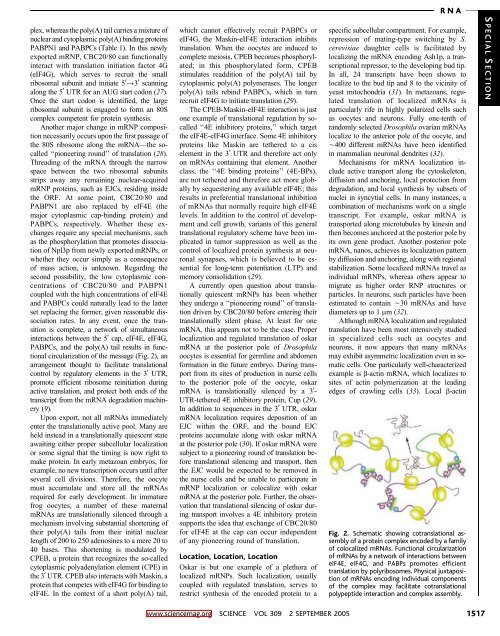
circularization of the message (Fig. 2), an<br />
arrangement thought to facilitate translational<br />
control by regulatory elements in the 3 UTR,<br />
promote efficient ribosome reinitiation during<br />
active translation, and protect both ends of the<br />
transcript from the mRNA degradation machinery<br />
(9).<br />
Upon export, not all mRNAs immediately<br />
enter the translationally active pool. Many are<br />
held instead in a translationally quiescent state<br />
awaiting either proper subcellular localization<br />
or some signal that the timing is now right to<br />
make protein. In early metazoan embryos, for<br />
example, no new transcription occurs until after<br />
several cell divisions. Therefore, the oocyte<br />
must accumulate and store all the mRNAs<br />
required for early development. In immature<br />
frog oocytes, a number of these maternal<br />
mRNAs are translationally silenced through a<br />
mechanism involving substantial shortening of<br />
their poly(A) tails from their initial nuclear<br />
length of 200 to 250 adenosines to a mere 20 to<br />
40 bases. This shortening is modulated by<br />
CPEB, a protein that recognizes the so-called<br />
cytoplasmic polyadenylation element (CPE) in<br />
the 3 UTR. CPEB also interacts with Maskin, a<br />
protein that competes with eIF4G for binding to<br />
eIF4E. In the context of a short poly(A) tail,<br />
which cannot effectively recruit PABPCs or<br />
eIF4G, the Maskin-eIF4E interaction inhibits<br />
translation. When the oocytes are induced to<br />
complete meiosis, CPEB becomes phosphorylated;<br />
in this phosphorylated form, CPEB<br />
stimulates readdition of the poly(A) tail by<br />
cytoplasmic poly(A) polymerases. The longer<br />
poly(A) tails rebind PABPCs, which in turn<br />
recruit eIF4G to initiate translation (29).<br />
The CPEB-Maskin-eIF4E interaction is just<br />
one example of translational regulation by socalled<br />
‘‘4E inhibitory proteins,’’ which target<br />
the eIF4E-eIF4G interface. Some 4E inhibitory<br />
proteins like Maskin are tethered to a cis<br />
element in the 3 UTR and therefore act only<br />
on mRNAs containing that element. Another<br />
class, the ‘‘4E binding proteins’’ (4E-BPs),<br />
are not tethered and therefore act more globally<br />
by sequestering any available eIF4E; this<br />
results in preferential translational inhibition<br />
of mRNAs that normally require high eIF4E<br />
levels. In addition to the control of development<br />
and cell growth, variants of this general<br />
translational regulatory scheme have been implicated<br />
in tumor suppression as well as the<br />
control of localized protein synthesis at neuronal<br />
synapses, which is believed to be essential<br />
for long-term potentiation (LTP) and<br />
memory consolidation (29).<br />
A currently open question about translationally<br />
quiescent mRNPs has been whether<br />
they undergo a ‘‘pioneering round’’ of translation<br />
driven by CBC20/80 before entering their<br />
translationally silent phase. At least for one<br />
mRNA, this appears not to be the case. Proper<br />
localization and regulated translation of oskar<br />
mRNA at the posterior pole of Drosophila<br />
oocytes is essential for germline and abdomen<br />
formation in the future embryo. During transport<br />
from its sites of production in nurse cells<br />
to the posterior pole of the oocyte, oskar<br />
mRNA is translationally silenced by a 3-<br />
UTR-tethered 4E inhibitory protein, Cup (29).<br />
In addition to sequences in the 3 UTR, oskar<br />
mRNA localization requires deposition of an<br />
EJC within the ORF, and the bound EJC<br />
proteins accumulate along with oskar mRNA<br />
at the posterior pole (30). If oskar mRNA were<br />
subject to a pioneering round of translation before<br />
translational silencing and transport, then<br />
theEJCwouldbeexpectedtoberemovedin<br />
the nurse cells and be unable to participate in<br />
mRNP localization or colocalize with oskar<br />
mRNA at the posterior pole. Further, the observation<br />
that translational silencing of oskar during<br />
transport involves a 4E inhibitory protein<br />
supports the idea that exchange of CBC20/80<br />
for eIF4E at the cap can occur independent<br />
of any pioneering round of translation.<br />
Location, Location, Location<br />
Oskar is but one example of a plethora of<br />
localized mRNPs. Such localization, usually<br />
coupled with regulated translation, serves to<br />
restrict synthesis of the encoded protein to a<br />
specific subcellular compartment. For example,<br />
repression of mating-type switching by S.<br />
cerevisiae daughter cells is facilitated by<br />
localizing the mRNA encoding Ash1p, a transcriptional<br />
repressor, to the developing bud tip.<br />
In all, 24 transcripts have been shown to<br />
localize to the bud tip and 8 to the vicinity of<br />
yeast mitochondria (31). In metazoans, regulated<br />
translation of localized mRNAs is<br />
particularly rife in highly polarized cells such<br />
as oocytes and neurons. Fully one-tenth of<br />
randomly selected Drosophila ovarian mRNAs<br />
localize to the anterior pole of the oocyte, and<br />
È400 different mRNAs have been identified<br />
in mammalian neuronal dendrites (32).<br />
Mechanisms for mRNA localization include<br />
active transport along the cytoskeleton,<br />
diffusion and anchoring, local protection from<br />
degradation, and local synthesis by subsets of<br />
nuclei in syncytial cells. In many instances, a<br />
combination of mechanisms work on a single<br />
transcript. For example, oskar mRNA is<br />
transported along microtubules by kinesin and<br />
then becomes anchored at the posterior pole by<br />
its own gene product. Another posterior pole<br />
mRNA, nanos, achieves its localization pattern<br />
by diffusion and anchoring, along with regional<br />
stabilization. Some localized mRNAs travel as<br />
individual mRNPs, whereas others appear to<br />
migrate as higher order RNP structures or<br />
particles. In neurons, such particles have been<br />
estimated to contain È30 mRNAs and have<br />
diameters up to 1 mm(32).<br />
Although mRNA localization and regulated<br />
translation have been most intensively studied<br />
in specialized cells such as oocytes and<br />
neurons, it now appears that many mRNAs<br />
may exhibit asymmetric localization even in somatic<br />
cells. One particularly well-characterized<br />
example is b-actin mRNA, which localizes to<br />
sites of actin polymerization at the leading<br />
edges of crawling cells (33). Local b-actin<br />
Fig. 2. Schematic showing cotranslational assembly<br />
of a protein complex encoded by a family<br />
of colocalized mRNAs. Functional circularization<br />
of mRNAs by a network of interactions between<br />
eIF4E, eIF4G, and PABPs promotes efficient<br />
translation by polyribosomes. Physical juxtaposition<br />
of mRNAs encoding individual components<br />
of the complex may facilitate cotranslational<br />
polypeptide interaction and complex assembly.<br />
S PECIAL S ECTION<br />
www.sciencemag.org SCIENCE VOL 309 2 SEPTEMBER 2005 1517