THIS WEEK IN
THIS WEEK IN
THIS WEEK IN

Create successful ePaper yourself
Turn your PDF publications into a flip-book with our unique Google optimized e-Paper software.
to an increased effort to improve methods for<br />
RNA crystallization (15). An encouraging sign<br />
was the appearance of the first crystal structures of a<br />
catalytic RNA, the hammerhead ribozyme, solved<br />
first as an RNA-DNA chimera and subsequently<br />
as an all-RNA structure (3, 4). Both structures<br />
revealed essentially the same fold, with three<br />
helices arranged in a Y configuration containing a<br />
U turn at the three-helix junction. Scott and coworkershavegoneontosolvethestructuresof<br />
four additional constructs by using strategies that<br />
trap the hammerhead ribozyme in different states<br />
of its catalytic cycle, revealing for the first time a<br />
detailed high-resolution ‘‘movie’’ of the mechanism<br />
of action of a catalytic RNA<br />
(16). Since the hammerhead structure,<br />
crystal structures of three more<br />
ribozymes have been solved, including<br />
the hepatitis delta virus ribozyme<br />
(17), the hairpin ribozyme (18), and<br />
the group I self-splicing intron<br />
(19–21), providing the structural<br />
basis for understanding their respective<br />
catalytic mechanisms.<br />
The first RNA structure to be<br />
solved that exceeded the size of<br />
tRNA was the 160-nt P4-P6 domain<br />
of the Tetrahymena group I intron<br />
at 2.8 ) resolution (5). It consists of<br />
two extended coaxial helical elements<br />
connected at one end by an<br />
internal loop containing a 150- bend<br />
(Fig. 1). For the first time, examples<br />
could be seen of the kinds of RNA-<br />
RNA interactions that are used to<br />
stabilize the packing of RNA helices<br />
into larger, more complex globular<br />
structures. One of these has been<br />
named the A-minor motif (22), one<br />
of the most abundant long-range<br />
interactions in rRNA, in which<br />
single-stranded adenosines make<br />
tertiary contacts with the minor<br />
grooves of double helices. A-minor<br />
interactions also play important<br />
functional roles. Helix-helix interactions<br />
were also formed by ribose<br />
zippers involving H bonding between<br />
the 2-hydroxyl group of a<br />
ribose in one helix and the 2-<br />
hydroxyl and the 2-oxygen of a<br />
pyrimidine base (or the 3-nitrogen of a purine<br />
base) of the other helix between their respective<br />
minor groove surfaces. In addition, close approach<br />
of phosphates was often mediated by<br />
bound hydrated magnesium ions. A recurring<br />
motif in the P4-P6 structure, called the A<br />
platform, positions adenines side by side in a<br />
pseudo–base pair within a helix, opening the<br />
minor groove for interactions with nucleotides<br />
from noncontiguous RNA strands.<br />
rRNA Secondary Structure Prediction<br />
Long before the first ribosome crystal structures<br />
appeared, the essential features of rRNA secondary<br />
structures were correctly predicted by<br />
using comparative sequence analysis (23–25).<br />
At about this same time, Michel and colleagues<br />
used a similar approach to establish the<br />
secondary structures of group I introns (26).<br />
Comparative analysis establishes base pairing<br />
by identification of compensating base changes<br />
in complementary nucleotides between two or<br />
more sequences. This approach was first explicitly<br />
applied by Fox and Woese (23), who,<br />
studying 5S rRNA sequences as phylogenetic<br />
markers, realized there was a common secondary<br />
structure that was compatible with several<br />
different sequences. Comparative analysis<br />
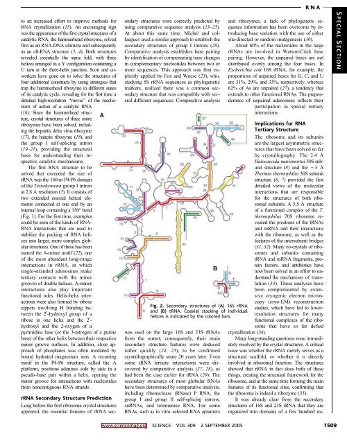
Fig. 2. Secondary structures of (A) 16S rRNA<br />
and (B) tRNA. Coaxial stacking of individual<br />
helices is indicated by the colored bars.<br />
wasusedonthelarge16Sand23SrRNAs<br />
from the outset; consequently, their main<br />
secondary structure features were deduced<br />
rather quickly (24, 25), to be confirmed<br />
crystallographically some 20 years later. Even<br />
some rRNA tertiary interactions were discovered<br />
by comparative analysis (27, 28), as<br />
hadbeenthecaseearlierfortRNA(29). The<br />
secondary structures of most globular RNAs<br />
have been determined by comparative analysis,<br />
including ribonuclease (RNase) P RNA, the<br />
group I and group II self-splicing introns,<br />
snRNAs, and telomerase RNA. For some<br />
RNAs, such as in vitro–selected RNA aptamers<br />
RNA<br />
and ribozymes, a lack of phylogenetic sequence<br />
information has been overcome by introducing<br />
base variation with the use of either<br />
site-directed or random mutagenesis (30).<br />
About 60% of the nucleotides in the large<br />
rRNAs are involved in Watson-Crick base<br />
pairing. However, the unpaired bases are not<br />
distributed evenly among the four bases. In<br />
Escherichia coli 16S rRNA, for example, the<br />
proportions of unpaired bases for G, C, and U<br />
are 31%, 29%, and 33%, respectively, whereas<br />
62% of As are unpaired (27), a tendency that<br />
extends to other functional RNAs. The preponderance<br />
of unpaired adenosines reflects their<br />
participation in special tertiary<br />
interactions.<br />
Implications for RNA<br />
Tertiary Structure<br />
The ribosome and its subunits<br />
are the largest asymmetric structures<br />
that have been solved so far<br />
by crystallography. The 2.4 )<br />
Halocarcula marismortui 50S subunit<br />
structure (8) andtheÈ3 )<br />
Thermus thermophilus 30S subunit<br />
structure (6, 7) provided the first<br />
detailed views of the molecular<br />
interactions that are responsible<br />
for the structures of both ribosomal<br />
subunits. A 5.5 ) structure<br />
of a functional complex of the T.<br />
thermophilus 70S ribosome revealed<br />
the positions of the tRNAs<br />
and mRNA and their interactions<br />
with the ribosome, as well as the<br />
features of the intersubunit bridges<br />
(31, 32). Many co-crystals of ribosomes<br />
and subunits containing<br />
tRNA and mRNA fragments, protein<br />
factors, and antibiotics have<br />
nowbeensolvedinanefforttounderstand<br />
the mechanism of translation<br />
(33). These analyses have<br />
been complemented by extensive<br />
cryogenic electron microscopy<br />
(cryo-EM) reconstruction<br />
studies, which have led to lowerresolution<br />
structures for many<br />
functional complexes of the ribosome<br />
that have so far defied<br />
crystallization (34).<br />
Many long-standing questions were immediately<br />
resolved by the crystal structures. A critical<br />
issue was whether the rRNA merely serves as a<br />
structural scaffold, or whether it is directly<br />
involved in ribosomal function. The structures<br />
showed that rRNA in fact does both of these<br />
things, creating the structural framework for the<br />
ribosome,andatthesametimeformingthemain<br />
features of its functional sites, confirming that<br />
the ribosome is indeed a ribozyme (35).<br />
It was already clear from the secondary<br />
structures of 16S and 23S rRNA that they are<br />
organized into domains of a few hundred nu-<br />
S PECIAL S ECTION<br />
www.sciencemag.org SCIENCE VOL 309 2 SEPTEMBER 2005 1509