in vitro culture and isoenzyme analysis of giardia lamblia
in vitro culture and isoenzyme analysis of giardia lamblia
in vitro culture and isoenzyme analysis of giardia lamblia
You also want an ePaper? Increase the reach of your titles
YUMPU automatically turns print PDFs into web optimized ePapers that Google loves.
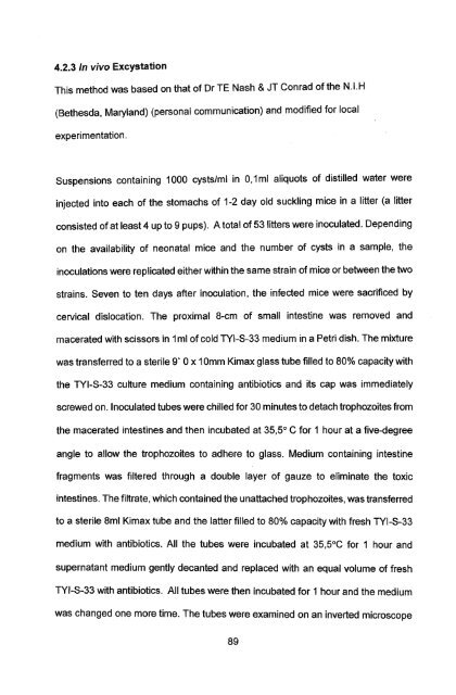
4.2.3 In vivo Excystation<br />
This method was based on that <strong>of</strong> Dr TE Nash & JT Conrad <strong>of</strong> the N.I.H<br />
(Bethesda, Maryl<strong>and</strong>) (personal communication) <strong>and</strong> modified for local<br />
experimentation.<br />
Suspensions conta<strong>in</strong><strong>in</strong>g 1000 cysts/m I <strong>in</strong> 0,1 ml aliquots <strong>of</strong> distilled water were<br />
<strong>in</strong>jected <strong>in</strong>to each <strong>of</strong> the stomachs <strong>of</strong> 1-2 day old suckl<strong>in</strong>g mice <strong>in</strong> a litter (a litter<br />
consisted <strong>of</strong> at least 4 up to 9 pups). A total <strong>of</strong> 53 litters were <strong>in</strong>oculated. Depend<strong>in</strong>g<br />
on the availability <strong>of</strong> neonatal mice <strong>and</strong> the number <strong>of</strong> cysts <strong>in</strong> a sample, the<br />
<strong>in</strong>oculations were replicated either with<strong>in</strong> the same stra<strong>in</strong> <strong>of</strong> mice or between the two<br />
stra<strong>in</strong>s. Seven to ten days after <strong>in</strong>oculation, the <strong>in</strong>fected mice were sacrificed by<br />
cervical dislocation. The proximal 8-cm <strong>of</strong> small <strong>in</strong>test<strong>in</strong>e was removed <strong>and</strong><br />
macerated with scissors <strong>in</strong> 1 ml <strong>of</strong> cold TYI-S-33 medium <strong>in</strong> a Petri dish. The mixture<br />
was transferred to a sterile 9' 0 x 10mm Kimax glass tube filled to 80% capacity with<br />
the TYI-S-33 <strong>culture</strong> medium conta<strong>in</strong><strong>in</strong>g antibiotics <strong>and</strong> its cap was immediately<br />
screwed on. Inoculated tubes were chilled for 30 m<strong>in</strong>utes to detach trophozoites from<br />
the macerated <strong>in</strong>test<strong>in</strong>es <strong>and</strong> then <strong>in</strong>cubated at 35,5° C for 1 hour at a five-degree<br />
angle to allow the trophozoites to adhere to glass. Medium conta<strong>in</strong><strong>in</strong>g <strong>in</strong>test<strong>in</strong>e<br />
fragments was filtered through a double layer <strong>of</strong> gauze to elim<strong>in</strong>ate the toxic<br />
<strong>in</strong>test<strong>in</strong>es. The filtrate, which conta<strong>in</strong>ed the unattached trophozoites, was transferred<br />
to a sterile 8ml Kimax tube <strong>and</strong> the latter filled to 80% capacity with fresh TYI-S-33<br />
medium with antibiotics. All the tubes were <strong>in</strong>cubated at 35,5°C for 1 hour <strong>and</strong><br />
supernatant medium gently decanted <strong>and</strong> replaced with an equal volume <strong>of</strong> fresh<br />
TYI-S-33 with antibiotics. All tubes were then <strong>in</strong>cubated for 1 hour <strong>and</strong> the medium<br />
was changed one more time. The tubes were exam<strong>in</strong>ed on an <strong>in</strong>verted microscope<br />
89


