in vitro culture and isoenzyme analysis of giardia lamblia
in vitro culture and isoenzyme analysis of giardia lamblia
in vitro culture and isoenzyme analysis of giardia lamblia

You also want an ePaper? Increase the reach of your titles
YUMPU automatically turns print PDFs into web optimized ePapers that Google loves.
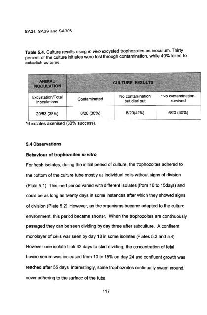
SA24 , SA29 <strong>and</strong> SA305.<br />
Table 5.4. Culture results us<strong>in</strong>g <strong>in</strong> vivo excysted trophozoites as <strong>in</strong>oculum. Thirty<br />
percent <strong>of</strong> the <strong>culture</strong> <strong>in</strong>itiates were lost through contam<strong>in</strong>ation, while 40% failed to<br />
establish <strong>culture</strong>s.<br />
ExcystationlT otal<br />
<strong>in</strong>oculations<br />
Contam<strong>in</strong>ated<br />
No contam<strong>in</strong>ation<br />
but died out<br />
*No contam<strong>in</strong>ationsurvived<br />
20/53 (38%)<br />
6/20 (30%)<br />
8/20(40%)<br />
6/20 (30%)<br />
*6 isolates axenised (30% success).<br />
5.4 Observations<br />
Behaviour <strong>of</strong> trophozoites <strong>in</strong> <strong>vitro</strong><br />
For fresh isolates, dur<strong>in</strong>g the <strong>in</strong>itial period <strong>of</strong> <strong>culture</strong>, the trophozoites adhered to<br />
the bottom <strong>of</strong> the <strong>culture</strong> tube mostly as <strong>in</strong>dividual cells without signs <strong>of</strong> division<br />
(Plate 5.1). This <strong>in</strong>ert period varied with different isolates (from 10 to 15days) <strong>and</strong><br />
could be as long as twenty days <strong>in</strong> some <strong>in</strong>stances after which they showed signs<br />
<strong>of</strong> division (Plate 5.2). However, as the organisms became adapted to the <strong>culture</strong><br />
environment, this period became shorter. When the trophozoites are cont<strong>in</strong>uously<br />
passaged they can be seen divid<strong>in</strong>g by day three after sub<strong>culture</strong>. A confluent<br />
monolayer <strong>of</strong> cells was seen by day 18 <strong>in</strong> some isolates (Plates 5.3 <strong>and</strong> 5.4)<br />
However one isolate took 32 days to start divid<strong>in</strong>g; the concentration <strong>of</strong> fetal<br />
bov<strong>in</strong>e serum was <strong>in</strong>creased from 10 to 15% on day 24 <strong>and</strong> confluent growth was<br />
reached after 55 days. Interest<strong>in</strong>gly, some trophozoites cont<strong>in</strong>ually swam around,<br />
never adher<strong>in</strong>g to the surface <strong>of</strong> the tube.<br />
117