

Vol 44 # 2 June 2012 - Kma.org.kw
Vol 44 # 2 June 2012 - Kma.org.kw
Vol 44 # 2 June 2012 - Kma.org.kw

Create successful ePaper yourself
Turn your PDF publications into a flip-book with our unique Google optimized e-Paper software.
93<br />
Proximal Tibial Osteotomy in Medial Compartment Osteoarthritis: How High is High?<br />
<strong>June</strong> <strong>2012</strong><br />
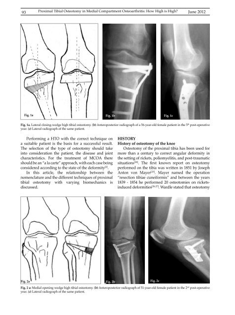
Fig. 1a Fig. 1b Fig. 1c<br />
Fig. 1a. Lateral closing-wedge high tibial osteotomy. (b) Anteroposterior radiograph of a 56-year-old female patient in the 5 th post-operative<br />
year. (c) Lateral radiograph of the same patient.<br />
Performing a HTO with the correct technique on<br />
a suitable patient is the basis for a successful result.<br />
The selection of the type of osteotomy should take<br />
into consideration the patient, the disease and joint<br />
characteristics. For the treatment of MCOA there<br />
should be an “a la carte” approach, with each case being<br />
considered according to the state of the deformity [2] .<br />
In this article, the relationship between the<br />
nomenclature and the different techniques of proximal<br />
tibial osteotomy with varying biomechanics is<br />
discussed.<br />
HISTORY<br />
History of osteotomy of the knee<br />
Osteotomy of the proximal tibia has been used for<br />
more than a century to correct angular deformity in<br />
the setting of rickets, poliomyelitis, and post-traumatic<br />
situations [24] . The first known report on osteotomy<br />
performed on the tibia was written in 1851 by Joseph<br />
Anton von Mayer [25] . Mayer named the operation<br />
“resection tibiae cuneiformis” and between the years<br />
1839 - 1854 he performed 20 osteotomies on ricketsinduced<br />
deformities [26,27] . Wardle stated that osteotomy<br />
Fig. 2a Fig. 2b Fig. 2c<br />
Fig. 2 a: Medial opening wedge high tibial osteotomy. (b) Anteroposterior radiograph of 51-year-old female patient in the 2 nd post-operative<br />
year. (c) Lateral radiograph of the same patient.


