

Vol 44 # 2 June 2012 - Kma.org.kw
Vol 44 # 2 June 2012 - Kma.org.kw
Vol 44 # 2 June 2012 - Kma.org.kw

Create successful ePaper yourself
Turn your PDF publications into a flip-book with our unique Google optimized e-Paper software.
<strong>June</strong> <strong>2012</strong><br />
KUWAIT MEDICAL JOURNAL 150<br />
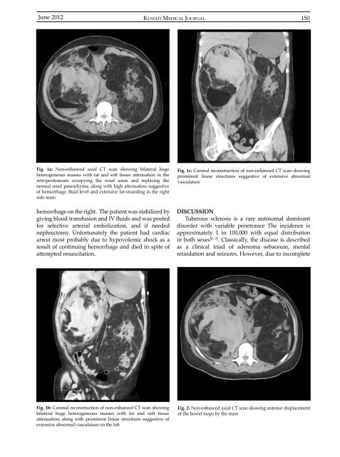
Fig. 1a: Non-enhanced axial CT scan showing bilateral huge<br />
heterogeneous masses with fat and soft tissue attenuation in the<br />
retroperitoneum occupying the renal areas and replacing the<br />
normal renal parenchyma; along with high attenuation suggestive<br />
of hemorrhage, fluid level and extensive fat-stranding in the right<br />
side mass<br />
hemorrhage on the right. The patient was stabilized by<br />
giving blood transfusion and IV fluids and was posted<br />
for selective arterial embolization, and if needed<br />
nephrectomy. Unfortunately the patient had cardiac<br />
arrest most probably due to hypovolemic shock as a<br />
result of continuing hemorrhage and died in spite of<br />
attempted resuscitation.<br />
Fig. 1c: Coronal reconstruction of non-enhanced CT scan showing<br />
prominent linear structures suggestive of extensive abnormal<br />
vasculature<br />
DISCUSSION<br />
Tuberous sclerosis is a rare autosomal dominant<br />
disorder with variable penetrance The incidence is<br />
approximately 1 in 100,000 with equal distribution<br />
in both sexes [8, 9] . Classically, the disease is described<br />
as a clinical triad of adenoma sebaceum, mental<br />
retardation and seizures. However, due to incomplete<br />
Fig. 1b: Coronal reconstruction of non-enhanced CT scan showing<br />
bilateral huge heterogeneous masses with fat and soft tissue<br />
attenuation; along with prominent linear structures suggestive of<br />
extensive abnormal vasculature on the left<br />
Fig. 2: Non-enhanced axial CT scan showing anterior displacement<br />
of the bowel loops by the mass


