

Vol 44 # 2 June 2012 - Kma.org.kw
Vol 44 # 2 June 2012 - Kma.org.kw
Vol 44 # 2 June 2012 - Kma.org.kw

Create successful ePaper yourself
Turn your PDF publications into a flip-book with our unique Google optimized e-Paper software.
<strong>June</strong> <strong>2012</strong><br />
KUWAIT MEDICAL JOURNAL 94<br />
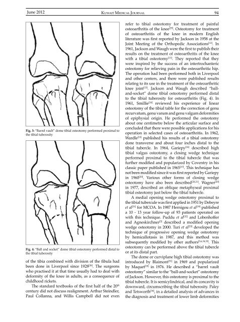
Fig. 3: “Barrel vault” dome tibial osteotomy performed proximal to<br />
the tibial tuberosity<br />
Fig. 4: “Ball and socket” dome tibial osteotomy performed distal to<br />
the tibial tuberosity<br />
of the tibia combined with division of the fibula had<br />
been done in Liverpool since 1928 [28] . The surgeons<br />
who practised it at that time usually had to deal with<br />
deformity of the knee in adults, as a consequence of<br />
childhood rickets.<br />
The standard textbooks of the first half of the 20 th<br />
century did not discuss realignment. Arthur Steindler,<br />
Paul Collanna, and Willis Campbell did not even<br />
refer to tibial osteotomy for treatment of painful<br />
osteoarthritis of the knee [29] . Osteotomy for treatment<br />
of osteoarthritis of the knee in modern English<br />
literature was first reported by Jackson in 1958 at the<br />
Joint Meeting of the Orthopedic Associations [12] . In<br />
1961, Jackson and Waugh were the first to publish their<br />
results on the treatment of osteoarthritis of the knee<br />
with a tibial osteotomy [13] . They reported that they<br />
were inspired by the success of an intertrochanteric<br />
osteotomy for relieving pain in the osteoarthritic hip.<br />
The operation had been performed both in Liverpool<br />
and other centers, and there were published results<br />
relating to its use in the treatment of the osteoarthritic<br />
knee joint [13] . Jackson and Waugh described “balland-socket”<br />
dome tibial osteotomy performed distal<br />
to the tibial tuberosity for osteoarthritis (Fig. 4). In<br />
1961, Smillie [14] reviewed his experience of linear<br />
osteotomy of the tibial table for the correction of genu<br />
recurvatum, genu varum and genu valgum deformities<br />
of epiphysial origin. He performed the osteotomy<br />
about one centimetre below the articular surface and<br />
concluded that there were possible applications for his<br />
operation in selected cases of osteoarthritis. In 1962,<br />
Wardle [15] published his results of a tibial osteotomy<br />
done transverse and about four inches distal to the<br />
tibial tubercle. In 1964, Gariepy [16] described high<br />
tibial valgus osteotomy, a closing wedge technique<br />
performed proximal to the tibial tubercle that was<br />
further modified and popularized by Coventry in his<br />
classic paper published in 1965 [17] . This technique has<br />
not been modified since it was first reported by Gariepy<br />
in 1960 [30] . Various other forms of closing wedge<br />
osteotomy have also been described [20,21] . Wagner [20]<br />
in 1977, described an oblique metaphyseal proximal<br />
tibial osteotomy just below the tibial tubercle.<br />
A medial opening wedge osteotomy proximal to<br />
the tibial tubercule was first applied in 1951 by Debeyre<br />
et al [31] for MCOA. In 1987 Hernigou et al [19] published<br />
a 10 - 13 year follow-up of 93 patients operated on<br />
with this technique. Puddu et al [32] and Lobenhoffer<br />
and Agneskirchner [3] described a modified opening<br />
wedge osteotomy in 2000. Turi et al [33] developed the<br />
technique of progressive opening wedge osteotomy<br />
by hemicallotasis in 1987, and this method was<br />
subsequently modified by other authors [5,6,34,35] . This<br />
osteotomy can be performed above the tibial tubercle<br />
or at its distal part.<br />
The dome or curviplane high tibial osteotomy was<br />
introduced by Blaimont [31] in 1969 and popularized<br />
by Maquet [18] in 1976. He described a “barrel vault<br />
osteotomy” similar to the “ball-and-socket” osteotomy<br />
of Jackson. However, this osteotomy is proximal to the<br />
tibial tubercle. It is semicylindrical, and its concavity is<br />
downward, circumscribing the tibial tuberosity. Paley<br />
and Tetsworth [36] , in a detailed analysis of advances in<br />
the diagnosis and treatment of lower limb deformities


